. 2010 Nov;225(2):454-65.
doi: 10.1002/jcp.22222.

Centrosome biogenesis continues in the absence of microtubules during prolonged S-phase arrest

Affiliations

Centrosome biogenesis continues in the absence of microtubules during prolonged S-phase arrest

Elizabeth S Collins et al. J Cell Physiol. 2010 Nov.

Abstract

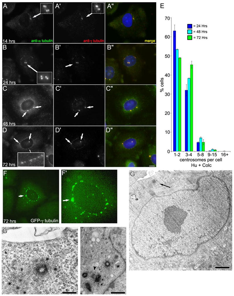
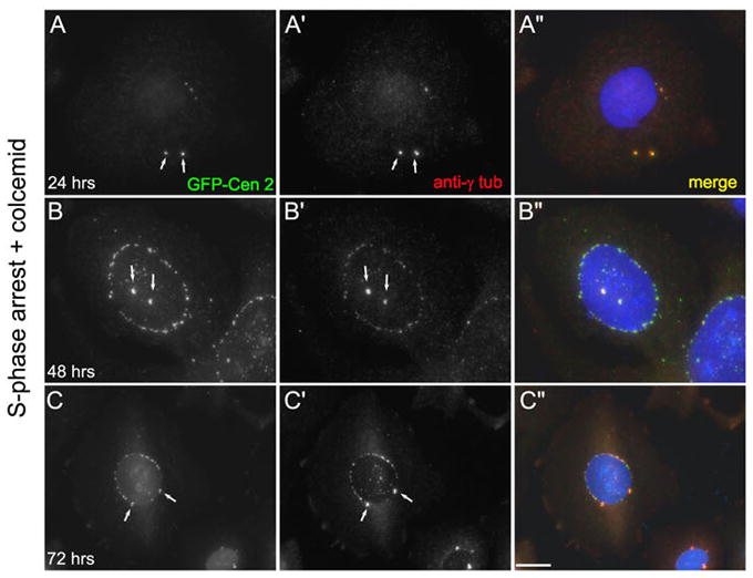
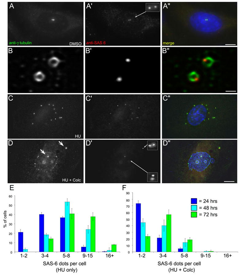
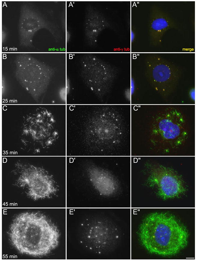
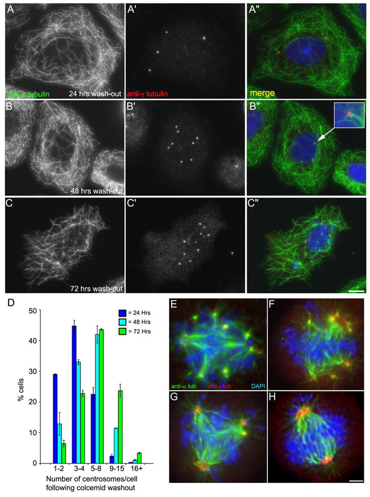
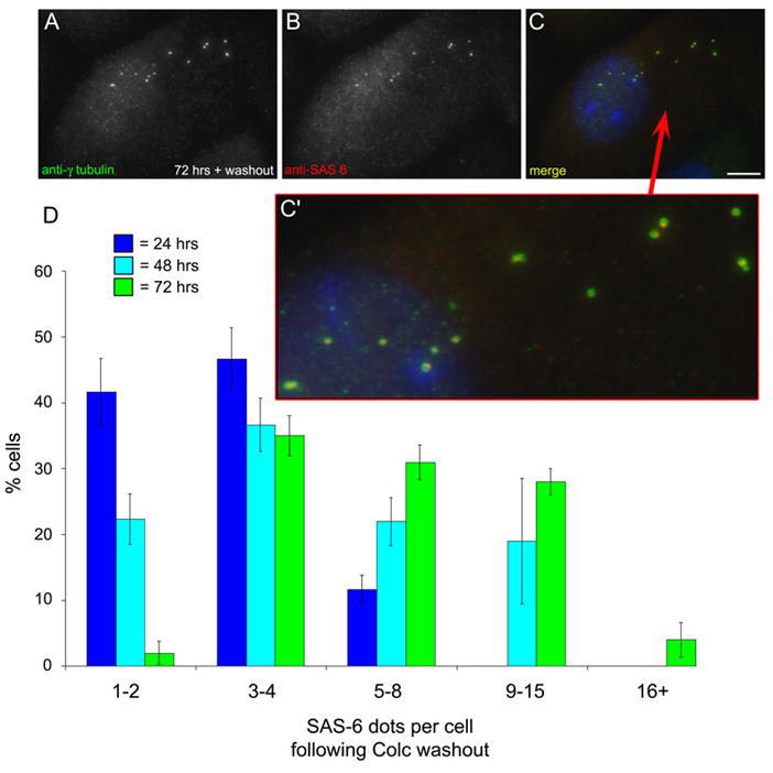
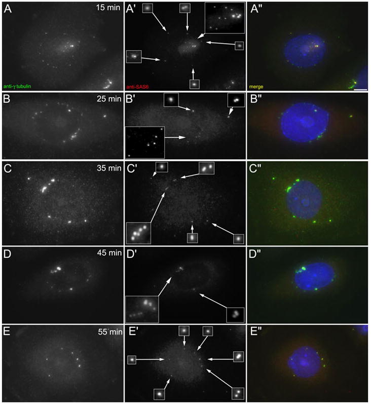
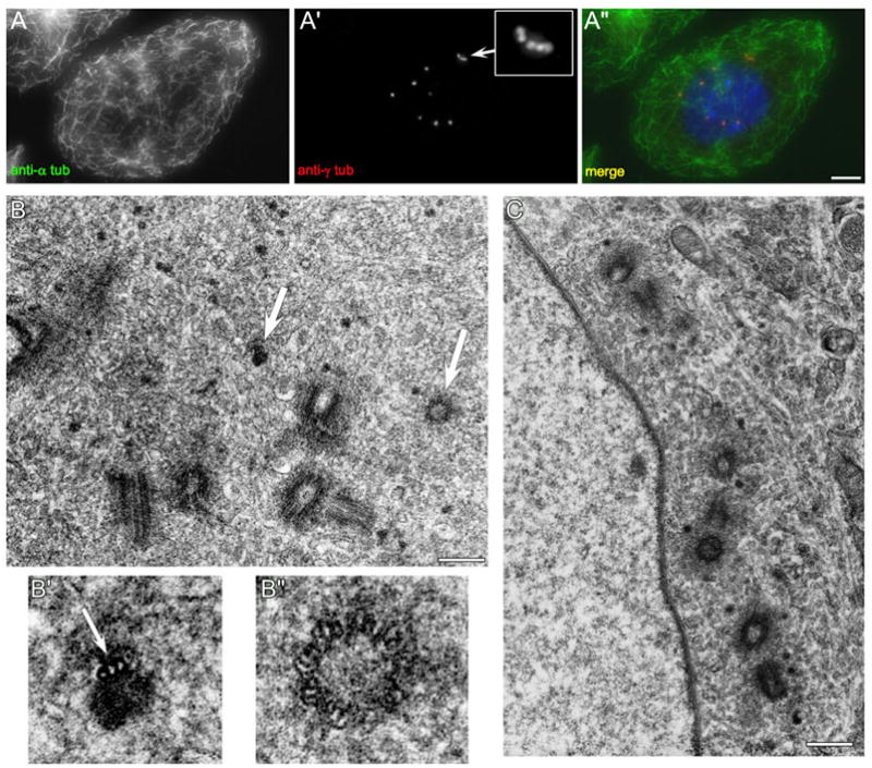
When CHO cells are arrested in S-phase, they undergo repeated rounds of centrosome duplication without cell-cycle progression. While the increase is slow and asynchronous, the number of centrosomes in these cells does rise with time. To investigate mechanisms controlling this duplication, we have arrested CHO cells in S-phase for up to 72 h, and coordinately inhibited new centriole formation by treatment with the microtubule poison colcemid. We find that in such cells, the pre-existing centrosomes remain, and a variable number of foci--containing alpha/gamma-tubulin and centrin 2--assemble at the nuclear periphery. When the colcemid is washed out, the nuclear-associated foci disappear, and cells assemble new centriole-containing centrosomes, which accumulate the centriole scaffold protein SAS-6, nucleate microtubule asters, and form functional mitotic spindle poles. The number of centrosomes that assemble following colcemid washout increases with duration of S-phase arrest, even though the number of nuclear-associated foci or pre-existing centrosomes does not increase. This suggests that during S-phase, a cryptic generative event occurs repeatedly, even in the absence of new triplet microtubule assembly. When triplet microtubule assembly is restored, these cryptic generative events become realized, and multiple centriole-containing centrosomes assemble.


Figures

Figure 1
Figure 1
Centrosome duplication does not continue in S phase arrested CHO-K1 cells treated with colcemid. AD: Immunofluorescent images of CHO-K1 cells labeled with anti-α-tubulin (A, B, C, D) and anti-γ-tubulin (A, B, C, D′). Cells were arrested in S phase for 12 hrs with hydroxyurea and then treated with hydroxyurea and colcemid for a further 2–60 hrs. Times given are from the initial addition of HU. A-A″: After 14 hrs, this cell contains two centrosomes represented by two γ-tubulin foci that colocalize with two α-tubulin foci, representing centrioles (inset). B-B″: After 24 h, the cell contains two centrosomes, each with a pair of centrioles (inset). Nuclear-associated foci, labeled with anti-α and γ-tubulin, are readily visible by this time point. CD: The irregular nuclear-associated foci persist at the 48 and 72 hr time points. The number of morphologically distinct centrosomes does not increase. E: Quantitation of the number of centrosomes per cell in S phase-arrested CHO-K1 cells treated with HU for 12 hrs, then HU/colc for 12, 36, or 60 hrs. Data is presented as the average number of centrosomes per cell +/− standard deviation for 24, 48, and 72 hrs. Note that the number of centrosomes does not substantially increase throughout the 72 hr time-course. F: Maximum intensity projections of S phase-arrested CHO-K1 cells stably transfected with γ-tubulin-GFP treated with colcemid. Note the irregular nuclear-associated foci are present in the perinuclear region, indicating that these foci are not an artifact of fixation. G: Transmission electron micrograph of a CHO-K1 cell arrested in S phase for 12 hrs with HU, followed by treatment with HU and colcemid for a further 60 hr. Only one centriole is present in this section - arrow (also see high-magnification view in G′). H: Transmission electron micrograph of a different cell from the same experiment showing four complete centrioles, and one pro-centriole (arrowhead). Scale bars: D = 10 μm, G = 0.5 μm, G″ = 0.1 μm, H = 0.2 μm.
Figure 2
Figure 2
Centrin 2 is a component of nuclear-associated foci. AC: Images of CHO-A8 cells, which stably express GFP-centrin 2, treated with HU and colcemid. A: At 24 hrs, the cell has two centrosomes (arrows) that are positive for centrin-2 and γ-tubulin. BC: At 48 and 72hrs, the cells have two centrosomes and numerous nuclear-associated centrosomal foci. Note the presence of irregular perinuclear foci positive for both γ-tubulin and centrin-2. Scale bar = 10 μm.
Figure 3
Figure 3
SAS-6 is recruited to the centrosomes, but not nuclear-associated foci in HU/colc-treated cells. AD: CHO cells labeled with antibodies against γ-tubulin and SAS-6. A-A″ shows an DMSO-control treated interphase cell with two γ-tubulin foci (centrosomes). Each foci has an associated SAS-6 positive foci (inset). B-B″ shows a higher magnification view of γ-tubulin/SAS-6 co-localization at the centrosome. The γ-tubulin is arrayed in a doughnut shape around the mature centriole, with a small focus of SAS-6 delineating the position of the nascent daughter centriole. C-C″ a CHO cell treated with HU for 72 hrs. This cell contains 12 γ-tubulin foci, each with an associated SAS-6 dot. D-D″ shows another CHO cell treated with HU/colc for 72 hrs. Here the cell has two prominent γ-tubulin foci (D, arrows), each with a pair of SAS-6 dots (D′, insets). Note that the nuclear associated foci in D are not recognized by the anit-SAS-6. Scale bars, A″ = 20 μm, B″ = 1 μm, C″ = 10 μm. E: CHO cells treated with HU for 24, 48, and 72 hrs stained with antibodies against SAS-6. The number of SAS-6 positive foci (dots) per cell was counted. Data is presented as the average of three experiments, +/− the standard deviation. F: CHO cells from parallel experiments, treated for 12 hrs with HU, followed by 12, 36, or 60 hrs of HU/colc. The number of SAS-6 dots per cell was counted, and the data presented as above. Note that the number of SAS-6 foci does not significantly increase over 72 hrs.
Figure 4
Figure 4
Time-course of colcemid washout in S-phase arrested CHO cells. AB: 15 and 25 min after colcemid removal, there are multiple γ-tubulin-containing foci around the nucleus and in the cytoplasm. Cytoplasmic microtubules remain absent. C: 35 min after colcemid removal, cytoplasmic microtubules are visible. There are multiple aster-like arrays, and these are γ-tubulin positive. DE: At 45 and 55 min after colcemid removal, the microtubule network continues to reform. The nuclear-associated foci have dispersed and are no longer visible. Instead, there are multiple γ-tubulin-positive microtubules asters. Scale bar = 10 μm.
Figure 5
Figure 5
The number of centrosomes that assemble following colcemid washout increases with the duration of cell cycle arrest. AC: Images of S phase-arrested CHO cells treated with colcemid, followed by colcemid removal 2 hrs prior to fixation. Cells are labeled with anti-α-tubulin and anti-γ-tubulin. A: After 2 hrs of colcemid washout, this cell has re-formed its microtubule network, and contains 4 distinct centrosomes. Note that the nuclear associated foci have dispersed. B: Colcemid washout after 48 hrs arrest, this cell has 8 distinct centrosomes. Note that these centrosomes lie in the center of the re-formed microtubule asters (inset). C: Washout after 72 hrs, the cell has 11 centrosomes. D: Distribution of the average number of centrosomes/cell following 2 hrs colcemid washout for 24, 48, and 72 hrs time points. Note the centrosome number increases with increase in the duration of cell cycle arrest. EH: Centrosomes formed after colcemid washout assemble functional spindle poles at mitosis. AD: Images of S phase-arrested CHO cells treated with HU and colcemid for 72 hrs. Both the colcemid and hydroxyurea were removed 12 hrs prior to fixation. Cells were stained with antiα-tubulin and anti-γ-tubulin. EF: Multiple spindle poles. G: A tripolar spindle with multiple centrosomes per pole. H: A bipolar spindle with multiple centrosomes per pole. Scale bar in C = 10 μm, H = 3 μm.
Figure 6
Figure 6
SAS-6 is recruited to multiple centrosomes following colcemid washout. AC′: CHO cells treated with HU for 12 hrs, followed by treatment with HU/colc for 60 hrs, then the colcemid removed for 2 hrs. Cells were fixed and stained with antibodies against γ-tubulin and SAS-6. The NAF have disassembled, and multiple centrosomes have assembled. Each is co-labeled with anti-SAS-6 (C′). Scale bar = 10 μm. D: The number of SAS-6 foci per cell was quantified for 2 hrs colcemid washout. The average number of SAS-6 positive centrosomes per cell has increased, compared to cells treated with HU/colc and no washout.
Figure 7
Figure 7
Rapid recruitment of SAS-6 to centrosomes following colcemid washout. AE: Immunofluorescent images of CHO cells treated with HU for 12 hrs, then HU/colc for a further 60 hrs. The colcemid was then washed out for the times indicated in each frame, and the cells fixed and immuno-labeled with antibodies against γ-tubulin and SAS-6. At T = 15 min (panels A-A″), the SAS-6 is recruited to multiple centrosomes, even before the NAF have disappeared. SAS-6 is localized to the major g-tubulin foci, but also to smaller foci. As the NAF disappear, and the g-tubulin foci become more distinct, these are positive for SAS-6 (C–E).
Figure 8
Figure 8
The centrosomes assembled after colcemid washout contain multiple centrioles. CHO cells were treated with HU for 12 hrs, then HU/colc for 60 hrs, followed by washout of colcemid for 2 hrs. A: 2 hrs after colcemid removal, the microtubule network is fully restored, and cells contain multiple centrosomes, including five clustered together (inset). B: These cells contain multiple centrioles. There are five complete centrioles in this section. In addition, there are two incomplete centrosome structures (arrows). B′: Magnified image of an electron dense focus associated with a single triplet microtubule. B″: Magnified image of a small, immature centriole. C: Another cell from the same preparation. There are seven complete centrioles visible in this section. Scale bars: A=10 μm, B, C = 0.4 μm.

Similar articles

Cited by

References

    1. Balczon R, Bao L, Zimmer W, Brown K, Zinkowski RP, Brinkley BR. Dissociation of centrosome replication events from cycles of DNA synthesis and mitotic division in hydroxyurea-arrested Chinese hamster ovary cells. J Cell Biol. 1995;130:105–115. - PMC - PubMed
    1. Balczon R, Varden CE, Schroer TA. Role for microtubules in centrosome doubling in Chinese hamster ovary cells. Cell Motil Cytoskeleton. 1999;42:60–72. - PubMed
    1. Basto R, Brunk K, Vinadogrova T, Peel N, Franz A, Khodjakov A, Raff JW. Centrosome amplification can initiate tumorigenesis in flies. Cell. 2008;133:1032–1042. - PMC - PubMed
    1. Bettencourt-Dias M, Glover DM. Centrosome biogenesis and function: centrosomics brings new understanding. Nat Rev Mol Cell Biol. 2007;8:451–463. - PubMed
    1. Bettencourt-Dias M, Rodrigues-Martins A, Carpenter L, Riparbelli M, Lehmann L, Gatt MK, Carmo N, Balloux F, Callaini G, Glover DM. SAK/PLK4 is required for centriole duplication and flagella development. Curr Biol. 2005;15:2199–2207. - PubMed

Publication types

LinkOut - more resources