

New fabrication techniques for ring-array transducers for real-time 3D intravascular ultrasound
- PMID: 20458877
- PMCID: PMC3057104
- DOI: 10.1177/016173460903100403
New fabrication techniques for ring-array transducers for real-time 3D intravascular ultrasound
Abstract
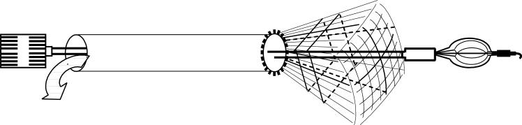
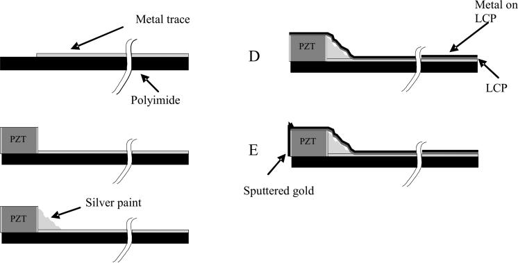
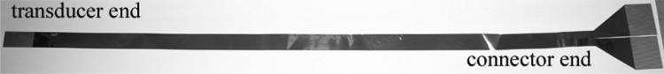
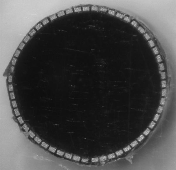
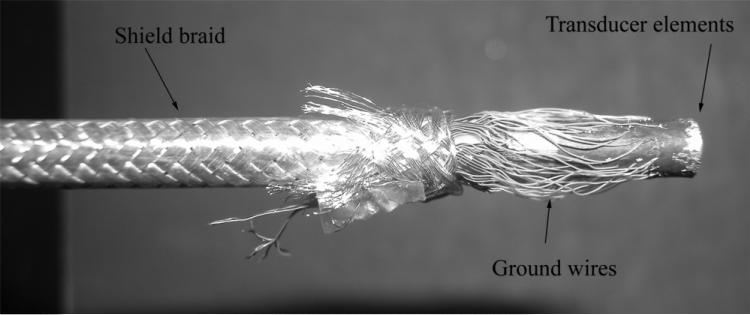
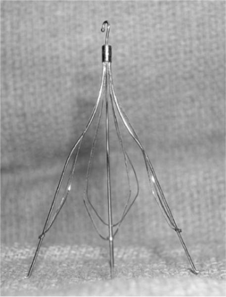
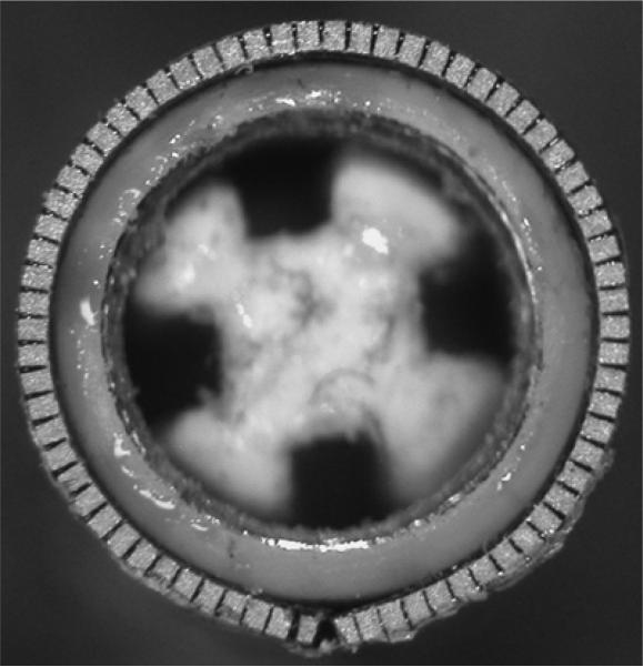
We have previously described miniature 2D array transducers integrated into a Cook Medical, Inc. vena cava filter deployment device. While functional, the fabrication technique was very labor intensive and did not lend itself well to efficient fabrication of large numbers of devices. We developed two new fabrication methods that we believe can be used to efficiently manufacture these types of devices in greater than prototype numbers. One transducer consisted of 55 elements operating near 5 MHz. The interelement spacing is 0.20 mm. It was constructed on a flat piece of copper-clad polyimide and then wrapped around an 11 French catheter of a Cook Medical, Inc. inferior vena cava (IVC) filter deployment device. We used a braided wiring technology from Tyco Electronics Corp. to connect the elements to our real-time 3D ultrasound scanner. Typical measured transducer element bandwidth was 20% centered at 4.7 MHz and the 50 Omega round trip insertion loss was --82 dB. The mean of the nearest neighbor cross talk was -37.0 dB. The second method consisted of a 46-cm long single layer flex circuit from MicroConnex that terminates in an interconnect that plugs directly into our system cable. This transducer had 70 elements at 0.157 mm interelement spacing operating at 4.8 MHz. Typical measured transducer element bandwidth was 29% and the 50 Omega round trip insertion loss was -83 dB. The mean of the nearest neighbor cross talk was -33.0 dB.
Figures

Similar articles
-
Progress in ring array transducers for real-time 3D ultrasound guidance of cardiac interventional devices.Ultrason Imaging. 2011 Jul;33(3):197-204. doi: 10.1177/016173461103300304. Ultrason Imaging. 2011. PMID: 21842583 Free PMC article.
-
Ring array transducers for real-time 3-D imaging of an atrial septal occluder.Ultrasound Med Biol. 2012 Aug;38(8):1483-7. doi: 10.1016/j.ultrasmedbio.2012.03.019. Epub 2012 Jun 12. Ultrasound Med Biol. 2012. PMID: 22698504 Free PMC article.
-
Real-time 3-D ultrasound guidance of interventional devices.IEEE Trans Ultrason Ferroelectr Freq Control. 2008 Sep;55(9):2066-78. doi: 10.1109/TUFFC.898. IEEE Trans Ultrason Ferroelectr Freq Control. 2008. PMID: 18986903 Free PMC article.
-
Intravascular ultrasound-guided bedside placement of inferior vena cava filters.Semin Vasc Surg. 2006 Sep;19(3):150-4. doi: 10.1053/j.semvascsurg.2006.06.002. Semin Vasc Surg. 2006. PMID: 16996417 Review.
-
Principles and devices.Semin Vasc Surg. 2006 Sep;19(3):128-31. doi: 10.1053/j.semvascsurg.2006.06.006. Semin Vasc Surg. 2006. PMID: 16996413 Review.
Cited by
-
Progress in ring array transducers for real-time 3D ultrasound guidance of cardiac interventional devices.Ultrason Imaging. 2011 Jul;33(3):197-204. doi: 10.1177/016173461103300304. Ultrason Imaging. 2011. PMID: 21842583 Free PMC article.
-
Ring array transducers for real-time 3-D imaging of an atrial septal occluder.Ultrasound Med Biol. 2012 Aug;38(8):1483-7. doi: 10.1016/j.ultrasmedbio.2012.03.019. Epub 2012 Jun 12. Ultrasound Med Biol. 2012. PMID: 22698504 Free PMC article.
-
Single-chip CMUT-on-CMOS front-end system for real-time volumetric IVUS and ICE imaging.IEEE Trans Ultrason Ferroelectr Freq Control. 2014 Feb;61(2):239-50. doi: 10.1109/TUFFC.2014.6722610. IEEE Trans Ultrason Ferroelectr Freq Control. 2014. PMID: 24474131 Free PMC article.
-
High-Frequency, 2-mm-Diameter Forward-Viewing 2-D Array for 3-D Intracoronary Blood Flow Imaging.IEEE Trans Ultrason Ferroelectr Freq Control. 2024 Aug;71(8):1051-1061. doi: 10.1109/TUFFC.2024.3418708. Epub 2024 Aug 19. IEEE Trans Ultrason Ferroelectr Freq Control. 2024. PMID: 38913530 Free PMC article.
-
Volumetric real-time imaging using a CMUT ring array.IEEE Trans Ultrason Ferroelectr Freq Control. 2012 Jun;59(6):1201-11. doi: 10.1109/TUFFC.2012.2310. IEEE Trans Ultrason Ferroelectr Freq Control. 2012. PMID: 22718870 Free PMC article.
References
-
- Eriksson M-O, Wanhainen A, Nyman R. Intravascular ultrasound with a vector phased-array probe (AcuNav) is feasible in endovascular abdominal aortic aneurysm repair. Acta Radiologica. 2009;50:870–875. - PubMed
-
- Light ED, Smith SW. Two dimensional arrays for real time 3D intravascular ultrasound. Ultrasonic Imaging. 2004;26:115–128. - PubMed
-
- Degertekin FL, Guldiken RO, Karaman M. Annular-ring CMUT arrays for forward-looking IVUS: transducer characterization and imaging. IEEE Trans Ultrason Ferroelec Freq Contr. 2006;53:474–482. - PubMed
-
- Yeh DT, Oralkan O, Wygant IO, O'Donnell M, Khuri-Yakub BT. 3-D ultrasound imaging using a forward-looking CMUT ring array for intravascular/intracardiac applications. IEEE Trans Ultrason Ferroelec Freq Contr. 2006;53:1202–1211. - PubMed
-
- Wang Y, Stephens DN, O'Donnell M. Optimizing the beam pattern of a forward-viewing ring-annular ultrasound array for intravascular imaging. IEEE Trans Ultrason Ferroelec Freq Contr. 2002;49:1652–1664. - PubMed
Publication types
MeSH terms
Grants and funding
LinkOut - more resources
Full Text Sources
Other Literature Sources
Miscellaneous
