

Electronic and protein structural dynamics of a photosensory histidine kinase
- PMID: 20459101
- PMCID: PMC2882989
- DOI: 10.1021/bi100527a
Electronic and protein structural dynamics of a photosensory histidine kinase
Abstract
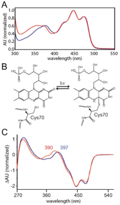
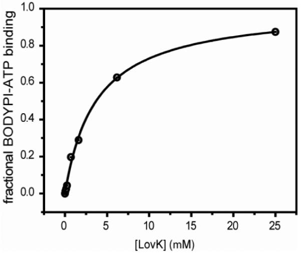
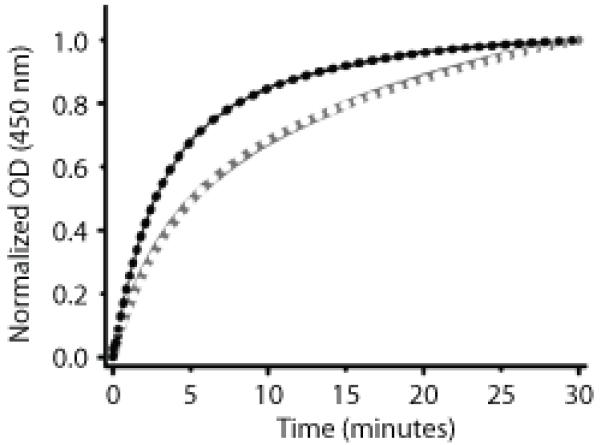
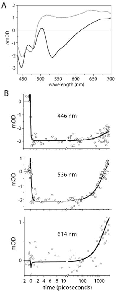
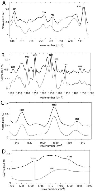
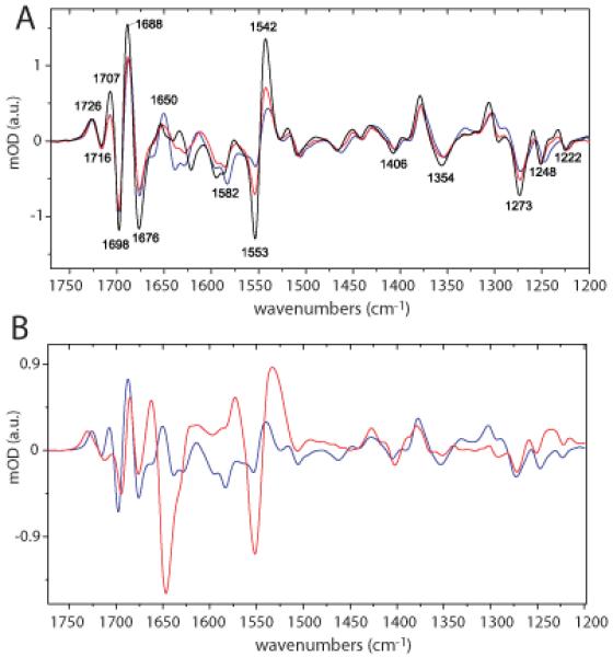
The bacterium Caulobacter crescentus encodes a two-component signaling protein, LovK, that contains an N-terminal photosensory LOV domain coupled to a C-terminal histidine kinase. LovK binds a flavin cofactor, undergoes a reversible photocycle, and displays regulated ATPase and autophosphorylation activity in response to visible light. Femtosecond to nanosecond visible absorption spectroscopy demonstrates congruence between full-length LovK and isolated LOV domains in the mechanism and kinetics of light-dependent cysteinyl-C4(a) adduct formation and rupture, while steady-state absorption and fluorescence line narrowing (FLN) spectroscopies reveal unique features in the electronic structure of the LovK flavin cofactor. In agreement with other sensor histidine kinases, ATP binds specifically to LovK with micromolar affinity. However, ATP binding to the histidine kinase domain of LovK has no apparent effect on global protein structure as assessed by differential Fourier transform infrared (FTIR) spectroscopy. Cysteinyl adduct formation results in only minor changes in the structure of LovK as determined by differential FTIR. This study provides insight into the structural underpinnings of LOV-mediated signal transduction in the context of a full-length histidine kinase. In particular, the data provide evidence for a model in which small changes in the tertiary/quaternary structure of LovK, as triggered by photon detection in the N-terminal LOV sensory domain, are sufficient to regulate histidine kinase activity.
Figures
References
-
- Hoch JA, Silhavy TJ. Two-Component Signal Transduction. ASM Press; Washington, D.C.: 1995.
-
- Crosson S, Rajagopal S, Moffat K. The LOV domain family: Photoresponsive signaling modules coupled to diverse output domains. Biochemistry. 2003;42:2–10. - PubMed
-
- Losi A. The bacterial counterparts of plant phototropins. Photochem Photobiol Sci. 2004;3:566–574. - PubMed
-
- Losi A. Flavin-based photoreceptors in bacteria. In: Silva E, Edwards AM, editors. Flavins - Photochemistry and Photobiology. RCS Publishing; Dorchester, UK: 2006. pp. 217–269.
-
- Losi A, Gärtner W. Bacterial bilin- and flavin-binding photoreceptors. Photochem Photobiol Sci. 2008;7:1168–1178. - PubMed
Publication types
MeSH terms
Substances
Grants and funding
LinkOut - more resources
Full Text Sources
Miscellaneous
