

3D critical plaque wall stress is a better predictor of carotid plaque rupture sites than flow shear stress: An in vivo MRI-based 3D FSI study
- PMID: 20459195
- PMCID: PMC2924579
- DOI: 10.1115/1.4001028
3D critical plaque wall stress is a better predictor of carotid plaque rupture sites than flow shear stress: An in vivo MRI-based 3D FSI study
Abstract
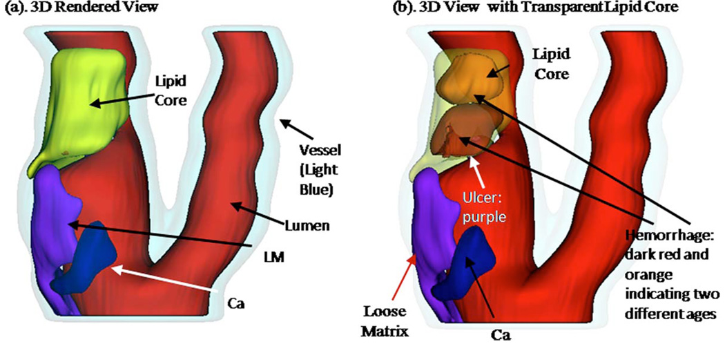
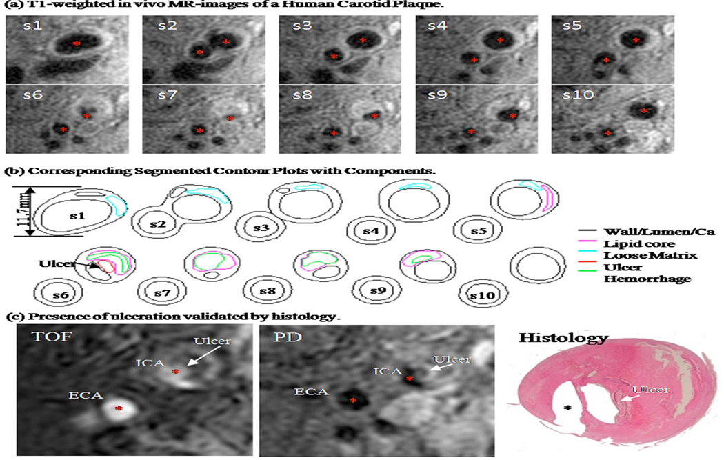
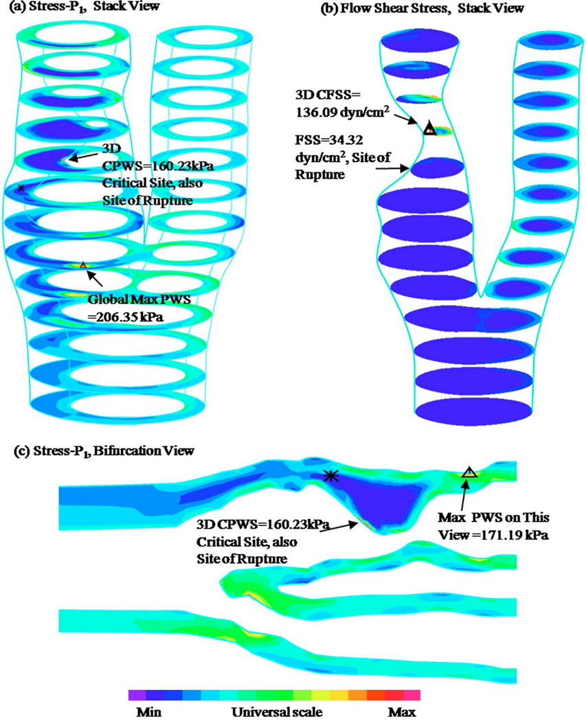
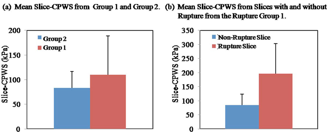
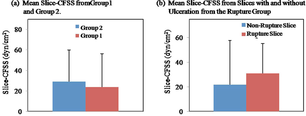
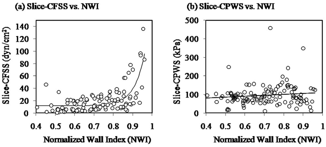
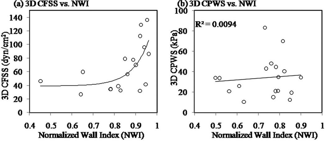
Atherosclerotic plaque rupture leading to stroke is the major cause of long-term disability as well as the third most common cause of mortality. Image-based computational models have been introduced seeking critical mechanical indicators, which may be used for plaque vulnerability assessment. This study extends the previous 2D critical stress concept to 3D by using in vivo magnetic resonance image (MRI) data of human atherosclerotic carotid plaques and 3D fluid-structure interaction (FSI) models to: identify 3D critical plaque wall stress (CPWS) and critical flow shear stress (CFSS) and to investigate their associations with plaque rupture. In vivo MRI data of carotid plaques from 18 patients scheduled for endarterectomy were acquired using histologically validated multicontrast protocols. Of the 18 plaques, histology-confirmed that six had prior rupture (group 1) as evidenced by presence of ulceration. The remaining 12 plaques (group 2) contained no rupture. The 3D multicomponent FSI models were constructed for each plaque to obtain 3D plaque wall stress (PWS) and flow shear stress (FSS) distributions. Three-dimensional CPWS and CFSS, defined as maxima of PWS and FSS from all vulnerable sites, were determined for each plaque to investigate their association with plaque rupture. Slice-based critical PWS and FSS were also calculated for all slices for more detailed analysis and comparison. The mean 3D CPWS of group 1 was 263.44 kPa, which was 100% higher than that from group 2 (132.77, p=0.03984). Five of the six ruptured plaques had 3D CPWS sites, matching the histology-confirmed rupture sites with an 83% agreement. Although the mean 3D CFSS (92.94 dyn/cm(2)) for group 1 was 76% higher than that for group 2 (52.70 dyn/cm(2)), slice-based CFSS showed no significant difference between the two groups. Only two of the six ruptured plaques had 3D CFSS sites matching the histology-confirmed rupture sites with a 33% agreement. CFSS had a good correlation with plaque stenosis severity (R(2)=0.40 with an exponential function fitting 3D CFSS data). This in vivo MRI pilot study using plaques with and without rupture demonstrates that 3D critical plaque wall stress values are more closely associated with atherosclerotic plaque rupture then critical flow shear stresses. Critical wall stress values may become indicators of high risk sites of rupture. Future work with a larger population will establish a possible CPWS-based plaque vulnerability classification.
Figures
Similar articles
-
Sites of rupture in human atherosclerotic carotid plaques are associated with high structural stresses: an in vivo MRI-based 3D fluid-structure interaction study.Stroke. 2009 Oct;40(10):3258-63. doi: 10.1161/STROKEAHA.109.558676. Epub 2009 Jul 23. Stroke. 2009. PMID: 19628799 Free PMC article.
-
Intraplaque hemorrhage is associated with higher structural stresses in human atherosclerotic plaques: an in vivo MRI-based 3D fluid-structure interaction study.Biomed Eng Online. 2010 Dec 31;9:86. doi: 10.1186/1475-925X-9-86. Biomed Eng Online. 2010. PMID: 21194481 Free PMC article.
-
3D MRI-based multicomponent FSI models for atherosclerotic plaques.Ann Biomed Eng. 2004 Jul;32(7):947-60. doi: 10.1023/b:abme.0000032457.10191.e0. Ann Biomed Eng. 2004. PMID: 15298432
-
MRI-based biomechanical parameters for carotid artery plaque vulnerability assessment.Thromb Haemost. 2016 Mar;115(3):493-500. doi: 10.1160/TH15-09-0712. Epub 2016 Jan 21. Thromb Haemost. 2016. PMID: 26791734 Review.
-
Systematic review of biomechanical forces associated with carotid plaque disruption and stroke.J Vasc Surg. 2025 May 14:S0741-5214(25)01043-2. doi: 10.1016/j.jvs.2025.05.014. Online ahead of print. J Vasc Surg. 2025. PMID: 40378930 Review.
Cited by
-
In vitro shear stress measurements using particle image velocimetry in a family of carotid artery models: effect of stenosis severity, plaque eccentricity, and ulceration.PLoS One. 2014 Jul 9;9(7):e98209. doi: 10.1371/journal.pone.0098209. eCollection 2014. PLoS One. 2014. PMID: 25007248 Free PMC article.
-
In vivo serial MRI-based models and statistical methods to quantify sensitivity and specificity of mechanical predictors for carotid plaque rupture: location and beyond.J Biomech Eng. 2011 Jun;133(6):064503. doi: 10.1115/1.4004189. J Biomech Eng. 2011. PMID: 21744932 Free PMC article.
-
Intraplaque stretch in carotid atherosclerotic plaque--an effective biomechanical predictor for subsequent cerebrovascular ischemic events.PLoS One. 2013 Apr 23;8(4):e61522. doi: 10.1371/journal.pone.0061522. Print 2013. PLoS One. 2013. PMID: 23626694 Free PMC article.
-
Rapid prediction of wall shear stress in stenosed coronary arteries based on deep learning.Front Bioeng Biotechnol. 2024 Aug 12;12:1360330. doi: 10.3389/fbioe.2024.1360330. eCollection 2024. Front Bioeng Biotechnol. 2024. PMID: 39188371 Free PMC article.
-
Quantifying effect of intraplaque hemorrhage on critical plaque wall stress in human atherosclerotic plaques using three-dimensional fluid-structure interaction models.J Biomech Eng. 2012 Dec;134(12):121004. doi: 10.1115/1.4007954. J Biomech Eng. 2012. PMID: 23363206 Free PMC article.
References
-
- Yuan C, Zhang SX, Polissar NL, Echelard D, Ortiz G, Davis JW, Ellington E, Ferguson MS, Hatsukami TS. Identification of Fibrous Cap Rupture With MRI Is Highly Associated With Recent Transient Ischemic Attack or Stroke. Circulation. 2002;105:181–185. - PubMed
-
- Casscells W, Naghavi M, Willerson JT. Vulnerable Atherosclerotic Plaque: A Multifocal Disease. Circulation. 2003;107:2072–2075. - PubMed
-
- Fuster V, Cornhill JF, Dinsmore RE, Fallon JT, Insull W, Libby P, Nissen S, Rosenfeld ME, Wagner WD, editors. AHA Monograph Series. Armonk, NY: Futura; 1998. The Vulnerable Atherosclerotic Plaque: Understanding, Identification, and Modification.
-
- Yuan C, Mitsumori LM, Beach KW, Maravilla KR. Special Review: Carotid Atherosclerotic Plaque: Noninvasive MR Characterization and Identification of Vulnerable Lesions. Radiology. 2001;221:285–299. - PubMed
-
- Saam T, Ferguson MS, Yarnykh VL, Takaya N, Xu D, Polissar NL, Hatsukami TS, Yuan C. Quantitative Evaluation of Carotid Plaque Composition by In Vivo MRI. Arterioscler., Thromb., Vasc., Biol. 2005;25(1):234–239. - PubMed
Publication types
MeSH terms
Grants and funding
LinkOut - more resources
Full Text Sources
Medical
