

doi: 10.1117/1.3369966.
Static and dynamic light scattering of healthy and malaria-parasite invaded red blood cells
Affiliations
- PMID: 20459219
- PMCID: PMC2862053
- DOI: 10.1117/1.3369966
Item in Clipboard
Static and dynamic light scattering of healthy and malaria-parasite invaded red blood cells
J Biomed Opt.
2010 Mar-Apr.
Abstract
We present the light scattering of individual Plasmodium falciparum-parasitized human red blood cells (Pf-RBCs), and demonstrate progressive alterations to the scattering signal arising from the development of malaria-inducing parasites. By selectively imaging the electric fields using quantitative phase microscopy and a Fourier transform light scattering technique, we calculate the light scattering maps of individual Pf-RBCs. We show that the onset and progression of pathological states of the Pf-RBCs can be clearly identified by the static scattering maps. Progressive changes to the biophysical properties of the Pf-RBC membrane are captured from dynamic light scattering.
Figures
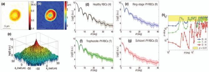
(a) Amplitude and (b) phase map of a healthy RBC. (c) The retrieved light scattering pattern of the same cell. Light-intensity scattering patterns of (d) healthy RBCs, (e) ring, (f) trophozoite, and (g) schizont stage of Pf-RBCs. (h) p-values of scattering patterns (different intraerythrocytic stages of Pf-RBCs are compared to the healthy RBCs).
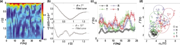
Dynamic light scattering of Pf-RBCs. (a) Normalized temporal autocorrelations of a healthy RBC as a function of decay time and scattering angle. (b) Normalized temporal autocorrelation of a healthy RBC at two representative scattering angles and fitted curves. (c) Line width Γ extracted from healthy and Pf RBCs as a function of scattering angle. Symbols represent the mean value and the error bars indicate the standard error for 15 RBCs. (d) Scatter plot of ω0 versus Γ taken for healthy and Pf RBCs. Each symbol represents mean values of 15 RBCs at scattering angles (0 to 21 deg). Lines indicate the boundaries of each group.
Similar articles
-
Light scattering of human red blood cells during metabolic remodeling of the membrane.J Biomed Opt. 2011 Jan-Feb;16(1):011013. doi: 10.1117/1.3524509. J Biomed Opt. 2011. PMID: 21280900 Free PMC article.
-
Synthetic Fourier transform light scattering.Opt Express. 2013 Sep 23;21(19):22453-63. doi: 10.1364/OE.21.022453. Opt Express. 2013. PMID: 24104134
-
Using elastic light scattering of red blood cells to detect infection of malaria parasite.IEEE Trans Biomed Eng. 2012 Jan;59(1):150-5. doi: 10.1109/TBME.2011.2168398. Epub 2011 Sep 15. IEEE Trans Biomed Eng. 2012. PMID: 21926010
-
The Plasmodium falciparum-infected red blood cell.Int J Biochem Cell Biol. 2011 Jun;43(6):839-42. doi: 10.1016/j.biocel.2011.03.012. Epub 2011 Mar 31. Int J Biochem Cell Biol. 2011. PMID: 21458590 Review.
-
The Rheopathobiology of Plasmodium vivax and Other Important Primate Malaria Parasites.Trends Parasitol. 2017 Apr;33(4):321-334. doi: 10.1016/j.pt.2016.11.009. Epub 2016 Dec 29. Trends Parasitol. 2017. PMID: 28040374 Review.
Cited by
-
Assessment of internal refractive index profile of stochastically inhomogeneous nuclear models via analysis of two-dimensional optical scattering patterns.J Biomed Opt. 2021 May;26(5):055001. doi: 10.1117/1.JBO.26.5.055001. J Biomed Opt. 2021. PMID: 33973424 Free PMC article.
-
In-depth biological analysis of alteration in Plasmodium knowlesi-infected red blood cells using a noninvasive optical imaging technique.Parasit Vectors. 2022 Mar 2;15(1):68. doi: 10.1186/s13071-022-05182-1. Parasit Vectors. 2022. PMID: 35236400 Free PMC article.
-
Light scattering of human red blood cells during metabolic remodeling of the membrane.J Biomed Opt. 2011 Jan-Feb;16(1):011013. doi: 10.1117/1.3524509. J Biomed Opt. 2011. PMID: 21280900 Free PMC article.
-
The Vanderbilt Memory & Aging Project: Study Design and Baseline Cohort Overview.J Alzheimers Dis. 2016 Mar 8;52(2):539-59. doi: 10.3233/JAD-150914. J Alzheimers Dis. 2016. PMID: 26967211 Free PMC article.
-
Matching an immersion medium's refractive index to a cell's cytosol isolates organelle scattering.Biomed Opt Express. 2022 Jul 19;13(8):4236-4246. doi: 10.1364/BOE.461874. eCollection 2022 Aug 1. Biomed Opt Express. 2022. PMID: 36032574 Free PMC article.
References
-
- Berne B. and Pecora R., Dynamic Light Scattering: with Applications to Chemistry, Biology, and Physics, Dover Publications, Mineola, New York: (2000).
-
- Subramanian H., Pradhan P., Liu Y., Capoglu I., Li X., Rogers J., Heifetz A., Kunte D., Roy H., Taflove A., and Backman V., “Optical methodology for detecting histologically unapparent nanoscale consequences of genetic alterations in biological cells,” Proc. Natl. Acad. Sci. U.S.A. PNASA6 105(51), 20118 (2008).10.1073/pnas.0804723105 - DOI - PMC - PubMed
Publication types
MeSH terms
Grants and funding
LinkOut - more resources
Full Text Sources
Other Literature Sources
