

doi: 10.1117/1.3339912.
Photoacoustic microscopy with 2-microm transverse resolution
Affiliations
- PMID: 20459224
- PMCID: PMC2857454
- DOI: 10.1117/1.3339912
Item in Clipboard
Photoacoustic microscopy with 2-microm transverse resolution
J Biomed Opt.
2010 Mar-Apr.
Abstract
We present a new-generation optical-resolution confocal photoacoustic microscope, consisting of a 0.25-numerical aperture optical microscope objective and a 75-MHz center-frequency spherically focused ultrasonic transducer. Experiments verified that this microscope has a transverse resolution of 2 microm, which is the highest to our knowledge among all photoacoustic imaging systems. In situ imaging of mouse ears shows the feasibility of resolving individual red blood cells in microvessels using the current system.
Figures

Schematic of the imaging probe in the 2-μm PAM system.
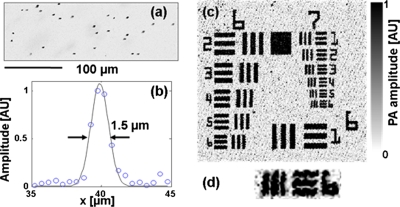
Experimental validation of transverse resolution of the 2-μm PAM system. (a) MAP image of a gelatin sample mixed with nanocages. (b) Transverse spread profile of a typical nanocage. Blue circle: experimental measurements. Black line: Gaussian-fit of the experimental data. (c) MAP image of a 1951 USAF resolution test target (groups 6 and 7). (d) Close-up view of MAP image of group 7 element 6 of the test target. (Color online only.)
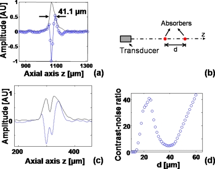
Axial resolution of the 2-μm PAM system. (a) Axial spread profile of a typical nanocage. Blue line with circle: experimental measurement. Black solid line: envelope of the experimental data. (b) Configuration of numerical shift-and-sum experiment. (c) Simulated photoacoustic signal and envelope when d=20 μm. Blue dashed line: photoacoustic signal. Black solid line: envelope of the signal. (d) CNR versus d. Blue circles: calculated CNR. Black dashed line: CNR=1. (Color online only.)

Microvasculature in mouse ear imaged using the 2-μm PAM system: (a) MAP image, (b) 3-D view of three representative x-y tomograms, and (c) close-up view of a typical dot in (a).

x-z cross-sectional photoacoustic image of two carbon fibers placed above and below a 300-μm-thick mouse ear (cf: carbon fibers. bv: blood vessels).

Animation showing how the measured photoacoustic signal and envelope evolve when the two absorbers move toward each other (MPEG, 1.05 MB). .

Animation shows the x-y tomograms of a mouse ear at different depth z (MPEG, 204 KB)..
References
-
- Wang L. V., “Tutorial on photoacoustic microscopy and computed tomography,” IEEE J. Sel. Top. Quant. 14(1), 171–179 (2008).10.1109/JSTQE.2007.913398 - DOI
Publication types
MeSH terms
Grants and funding
LinkOut - more resources
Full Text Sources
Medical
