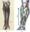
Real-time optoacoustic monitoring and three-dimensional mapping of a human arm vasculature
- PMID: 20459227
- PMCID: PMC2859082
- DOI: 10.1117/1.3370336
Real-time optoacoustic monitoring and three-dimensional mapping of a human arm vasculature
Abstract
We present our findings from a real-time laser optoacoustic imaging system (LOIS). The system utilizes a Q-switched Nd:YAG laser; a standard 128-channel ultrasonic linear array probe; custom electronics and custom software to collect, process, and display optoacoustic (OA) images at 10 Hz. We propose that this system be used during preoperative mapping of forearm vessels for hemodialysis treatment. To demonstrate the real-time imaging capabilities of the system, we show OA images of forearm vessels in a volunteer and compare our results to ultrasound images of the same region. Our OA images show blood vessels in high contrast. Manipulations with the probe enable us to locate and track arteries and veins of a forearm in real time. We also demonstrate the ability to combine a series of OA image slices into a volume for spatial representation of the vascular network. Finally, we use frame-by-frame analysis of the recorded OA video to measure dynamic changes of the crossection of the ulnar artery.
Figures


Similar articles
-
Optoacoustic imaging of the prostate: development toward image-guided biopsy.J Biomed Opt. 2010 Mar-Apr;15(2):021310. doi: 10.1117/1.3333548. J Biomed Opt. 2010. PMID: 20459232 Free PMC article.
-
Transverse flow imaging based on photoacoustic Doppler bandwidth broadening.J Biomed Opt. 2010 Mar-Apr;15(2):021304. doi: 10.1117/1.3339953. J Biomed Opt. 2010. PMID: 20459226 Free PMC article.
-
Real-time full-field photoacoustic imaging using an ultrasonic camera.J Biomed Opt. 2010 Mar-Apr;15(2):021318. doi: 10.1117/1.3420079. J Biomed Opt. 2010. PMID: 20459240
-
Laser optoacoustic imaging system for detection of breast cancer.J Biomed Opt. 2009 Mar-Apr;14(2):024007. doi: 10.1117/1.3086616. J Biomed Opt. 2009. PMID: 19405737
-
Doppler velocity measurements from large and small arteries of mice.Am J Physiol Heart Circ Physiol. 2011 Aug;301(2):H269-78. doi: 10.1152/ajpheart.00320.2011. Epub 2011 May 13. Am J Physiol Heart Circ Physiol. 2011. PMID: 21572013 Free PMC article. Review.
Cited by
-
Effect of irradiation distance on image contrast in epi-optoacoustic imaging of human volunteers.Biomed Opt Express. 2014 Oct 1;5(11):3765-80. doi: 10.1364/BOE.5.003765. eCollection 2014 Nov 1. Biomed Opt Express. 2014. PMID: 25426309 Free PMC article.
-
Handheld photoacoustic tomography probe built using optical-fiber parallel acoustic delay lines.J Biomed Opt. 2014 Aug;19(8):086007. doi: 10.1117/1.JBO.19.8.086007. J Biomed Opt. 2014. PMID: 25104413 Free PMC article.
-
Subwavelength-resolution label-free photoacoustic microscopy of optical absorption in vivo.Opt Lett. 2010 Oct 1;35(19):3195-7. doi: 10.1364/OL.35.003195. Opt Lett. 2010. PMID: 20890331 Free PMC article.
-
In vivo optoacoustic temperature imaging for image-guided cryotherapy of prostate cancer.Phys Med Biol. 2018 Mar 21;63(6):064002. doi: 10.1088/1361-6560/aab241. Phys Med Biol. 2018. PMID: 29480808 Free PMC article.
-
Photoacoustic/ultrasound dual imaging of human thyroid cancers: an initial clinical study.Biomed Opt Express. 2017 Jun 26;8(7):3449-3457. doi: 10.1364/BOE.8.003449. eCollection 2017 Jul 1. Biomed Opt Express. 2017. PMID: 28717580 Free PMC article.
References
-
- Oraevsky A. A., Jacques S. L., and Tittel F. K., “Determination of tissue optical properties by piezoelectric detection of laser-induced stress waves,” Proc. SPIE PSISDG 1882, 86–101 (1993).10.1117/12.147694 - DOI
-
- Oraevsky A. A., Jacques S. L., Esenaliev R. O., and Tittel F. K., “Time-resolved optoacoustic imaging in layered biological tissues,” in Advances in Optical Imaging, Alfano R. R., Ed., pp. 161–165, Academic Press, New York: (1994).
-
- Oraevsky A. A. and Karabutov A. A., “Optoacoustic tomography,” in Biomedical Photonics Handbook, Vo-Dinh T., Ed., pp. 34/31–34/34, CRC Press, Boca Raton, FL: (2003).
-
- Wang L. V., Photoacoustic Imaging and Spectroscopy, CRC Press, New York: (2009).
Publication types
MeSH terms
Grants and funding
LinkOut - more resources
Full Text Sources
Other Literature Sources

