. 2010 Mar-Apr;15(2):021310.
doi: 10.1117/1.3333548.

Optoacoustic imaging of the prostate: development toward image-guided biopsy

Affiliations

Optoacoustic imaging of the prostate: development toward image-guided biopsy

Mohammad A Yaseen et al. J Biomed Opt. 2010 Mar-Apr.

Abstract

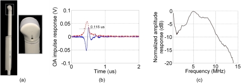
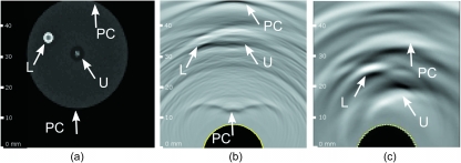
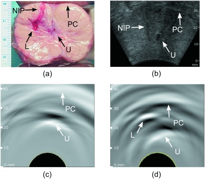
Optoacoustic (OA) tomography has demonstrated utility in identifying blood-rich malignancies in breast tissue. We describe the development and characterization of a laser OA imaging system for the prostate (LOIS-P). The system consists of a fiber-coupled Q-switched laser operating at 757 nm, a commercial 128-channel ultrasonic probe, a digital signal processor, and software that uses the filtered radial back-projection algorithm for image reconstruction. The system is used to reconstruct OA images of a blood-rich lesion induced in vivo in a canine prostate. OA images obtained in vivo are compared to images acquired using ultrasound, the current gold standard for guiding biopsy of the prostate. Although key structural features such as the urethra could be identified with both imaging techniques, a bloody lesion representing a highly vascularized tumor could only be clearly identified in OA images. The advantages and limitations of both forward and backward illumination modes are also evaluated by collecting OA images of phantoms simulating blood vessels within tissue. System resolution is estimated to be 0.2 mm in the radial direction of the acoustic array. The minimum detectable pressure signal is 1.83 Pa. Our results encourage further development toward a dual-modality OA/ultrasonic system for prostate imaging and image-guided biopsy.


Figures

Figure 1
Figure 1
(a) Commercial 128-channel endocavity ultrasound probe (EC7ART, Scanhead Corp., San Jose, California) used to collect acoustic transients for OA imaging. The piezoceramic detectors are oriented as a convex array lining the edge of the probe, as seen in the enlarged section. (b) Measured OA impulse response of a single detector in the endocavity probe, where the dashed line indicates envelope-detected signal used for estimation of the radial resolution (see text) and (c) the corresponding frequency response.
Figure 2
Figure 2
Images of combined OA signals for 1-mm cylindrical tube (with the tube axis oriented orthogonal to the imaging plane) positioned at various depths within the aqueous medium simulating optical properties of background tissues, and illuminated using (a) backward OA mode and (b) forward OA mode. OA signals were acquired using a single target, and the images were subsequently created after combining the signals from each individual acquisition. The images are displayed using the standard 8-bit gray-scale palette. The axes are scaled in millimeters.
Figure 3
Figure 3
Comparison of the modeled OA image with the image acquired in vivo: (a) imaging slice showing the distribution of the absorbed optical energy in a prostate model with spherical malignant lesion, determined by MC simulation; (b) OA image constructed using the simulated acoustic transients and the RBP algorithm; and (c) OA image of the dog prostate obtained in vivo after inducing a blood-rich lesion. The prostate capsule can be delineated in the OA images. The urethra and the lesion can be seen in the OA images as a dark and bright spots, respectively. The absorption difference between the lesion and surrounding tissue results in high contrast of the OA image. The low absorption in the urethra yields low OA signals. On each image, arrows indicate the prostate capsule (PC), urethra (U), and lesion (L). The images are displayed using the standard 8-bit gray-scale palette.
Figure 4
Figure 4
(a) Photograph of the sliced dog prostate showing the presence of the induced lesion with blood in the right lateral lobe, the urethra is visible but contracted after surgical excision; (b) ultrasonic image of the same dog prostate obtained in vivo after the surgery. OA images of the same dog prostate obtained in vivo (c) before and (d) after the lesion was induced. The induced bloody lesion can be seen in (a) and (d). The needle insertion path is visible in (a) and (b). Due to the acoustic mismatch between tissue and air, the prostate capsule can be identified in OA images as a white band. Arrows indicate the prostate capsule (PC), urethra (U), needle insertion path (NIP), and lesion (L). The images are displayed using the standard 8-bit gray-scale palette.

Similar articles

Cited by

References

    1. Cancer Facts & Figures 2009, American Cancer Society, Atlanta: (2009).
    1. Jemal A., Siegel R., Ward E., Hao Y., Xu J., and Thun M. J., “Cancer statistics, 2009,” Ca-Cancer J. Clin. CAMCAM 59, 225–249 (2009).10.3322/caac.20006 - DOI - PubMed
    1. Bracarda S., Cobelli O. d., Greco C., Prayer-Galetti T., Valdagni R., Gatta G., Braud F. d., and Bartsch G., “Cancer of the prostate,” Crit. Rev. Oncol. Hematol. ZZZZZZ 56, 379–396 (2005).10.1016/j.critrevonc.2005.03.010 - DOI - PubMed
    1. Wink M. H., Rosette J. J. M. M. C. d. l., Grimbergen C. A., and Wijkstra H., “Transrectal contrast enhanced ultrasound for diagnosis of prostate cancer,” World J. Urol. WJURDJ 25, 367–373 (2007).10.1007/s00345-007-0189-0 - DOI - PubMed
    1. Aarnink R. G., Beerlage H. P., Rosette J. J. M. C. H. D. L., Debruyne F. M. J., and Wijkstra H., “Transrectal ultrasound of the prostate: innovations and future applications,” J. Urol. 159, 1568–1579 (1998).10.1097/00005392-199805000-00045 - DOI - PubMed

Publication types