. 2010 Mar-Apr;15(2):021316.
doi: 10.1117/1.3365937.

Silver nanosystems for photoacoustic imaging and image-guided therapy

Affiliations

Silver nanosystems for photoacoustic imaging and image-guided therapy

Kimberly Homan et al. J Biomed Opt. 2010 Mar-Apr.

Abstract

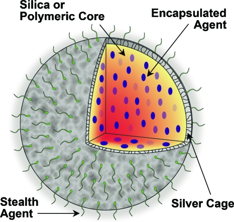
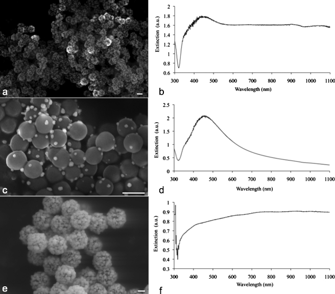
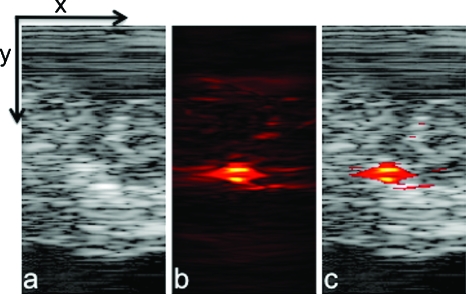
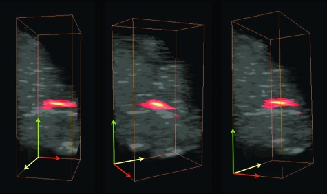
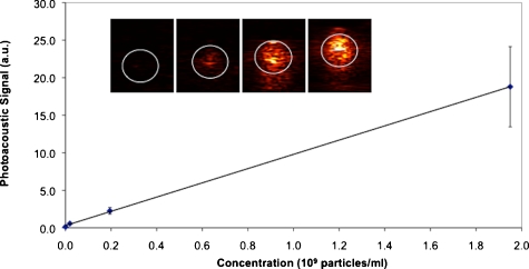
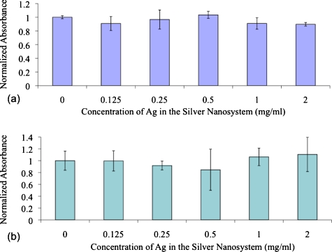
Due to their optical absorption properties, metallic nanoparticles are excellent photoacoustic imaging contrast agents. A silver nanosystem is presented here as a potential contrast agent for photoacoustic imaging and image-guided therapy. Currently, the nanosystem consists of a porous silver layer deposited on the surface of spherical silica cores ranging in diameter from 180 to 520 nm. The porous nature of the silver layer will allow for release of drugs or other therapeutic agents encapsulated in the core in future applications. In their current PEGylated form, the silver nanosystem is shown to be nontoxic in vitro at concentrations of silver up to 2 mgml. Furthermore, the near-infrared absorbance properties of the nanosystem are demonstrated by measuring strong, concentration-dependent photoacoustic signal from the silver nanosystem embedded in an ex vivo tissue sample. Our study suggests that silver nanosystems can be used as multifunctional agents capable of augmenting image-guided therapy techniques.


Figures

Figure 1
Figure 1
Multifunctional nanosystem platform capable of providing imaging contrast, drug delivery, and image-guided therapy.
Figure 2
Figure 2
Schematic of the combined photoacoustic and ultrasound (PAUS) imaging system incorporating the array-based ultrasound transducer integrated with the fiber optical light delivery system.
Figure 3
Figure 3
(a) Scanning electron micrograph (SEM) of the silver nanosystem with silica core diameter of 180 nm, and (b) corresponding coated 180-nm UV-vis spectrograph; (c) a batch of sparsely coated 180-nm nanoparticles, and (d) corresponding UV-vis spectrograph; (e) a silver-coated 520-nm silica core batch, and (f) corresponding UV-vis spectrograph. All scale bars are 200 nm.
Figure 4
Figure 4
(a) Ultrasound, (b) photoacoustic, and (c) combined images of nanoparticles injected directly into an ex vivo canine pancreas. All images were acquired from the same position as determined by the location of the ultrasound transducer. The images are 20 mm deep (y axis) and 10.5 mm wide (x axis).
Figure 5
Figure 5
Several views of a 3-D rendering of PAUS imaging from the silver nanosystem injected directly into an ex vivo canine pancreas. All images are 20×10.5×12 mm. For clarity, ultrasound signal from gelatin above the tissue was suppressed in these images.
Figure 6
Figure 6
Plot of fluence-normalized photoacoustic signal versus nanoparticle concentration for the four samples shown in the inset. Inset: photoacoustic images of gelatin phantom with PVA samples containing 0, 2×10⁷, 2×10⁸, and 2×10⁹ particles per ml from left to right, where the white circle outlines the boundaries of the samples as determined by ultrasound imaging. All inset images are 12.5×10.5 mm.
Figure 7
Figure 7
Cell viability data after 24-h exposure to the silver nanosystem at different concentrations in (a) MDA-MB-231 cells and (b) MPanc96 cells.
Video 1
Video 1
A movie of the 3-D rendering of PAUS imaging from the silver nanosystem injected directly into an ex vivo canine pancreas. The image covers a 20×10.5×12-mm volume. For clarity, ultrasound signal from gelatin above the tissue was suppressed (QuickTime, 1.97 MB). .

References

    1. Chen H., Roco M. C., Li X., and Lin Y., “Trends in nanotechnology patents,” Nat. Nanotechnol. NNAABX 3(3), 123–125 (2008).10.1038/nnano.2008.51 - DOI - PubMed
    1. Roco M. C., “Nanotechnology: convergence with modern biology and medicine,” Curr. Opin. Biotechnol. CUOBE3 14(3), 337–346 (2003).10.1016/S0958-1669(03)00068-5 - DOI - PubMed
    1. Bell A. G., “Production of sound by radiant energy,” J. Franklin Inst. JFINAB 111(6), 401–426 (1881).10.1016/0016-0032(81)90005-3 - DOI
    1. Emelianov S., Aglyamov S., Shah J., Sethuraman S., Scott G., Schmitt R., Karpiouk A., Motamedi M., and Oraevsky A., “Synergy of ultrasound, elasticity, and optoacoustic imaging for improved detection and differentiation of cancerous tissue,” J. Acoust. Soc. Am. JASMAN 115, 2411 (2004).10.1121/1.1621858 - DOI
    1. Emelianov S. Y., Aglyamov S. R., Shah J., Sethuraman S., Scott W. G., Schmitt R., Motamedi M., Karpiouk A., and Oraevsky A., “Combined ultrasound, optoacoustic, and elasticity imaging,” Proc. SPIE PSISDG 5320, 101–112 (2004).10.1117/12.537155 - DOI

Publication types