Evaluation of quantitative image analysis criteria for the high-resolution microendoscopic detection of neoplasia in Barrett's esophagus
- PMID: 20459272
- PMCID: PMC2874050
- DOI: 10.1117/1.3406386
Evaluation of quantitative image analysis criteria for the high-resolution microendoscopic detection of neoplasia in Barrett's esophagus
Abstract
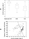
Early detection of neoplasia in patients with Barrett's esophagus is essential to improve outcomes. The aim of this ex vivo study was to evaluate the ability of high-resolution microendoscopic imaging and quantitative image analysis to identify neoplastic lesions in patients with Barrett's esophagus. Nine patients with pathologically confirmed Barrett's esophagus underwent endoscopic examination with biopsies or endoscopic mucosal resection. Resected fresh tissue was imaged with fiber bundle microendoscopy; images were analyzed by visual interpretation or by quantitative image analysis to predict whether the imaged sites were non-neoplastic or neoplastic. The best performing pair of quantitative features were chosen based on their ability to correctly classify the data into the two groups. Predictions were compared to the gold standard of histopathology. Subjective analysis of the images by expert clinicians achieved average sensitivity and specificity of 87% and 61%, respectively. The best performing quantitative classification algorithm relied on two image textural features and achieved a sensitivity and specificity of 87% and 85%, respectively. This ex vivo pilot trial demonstrates that quantitative analysis of images obtained with a simple microendoscope system can distinguish neoplasia in Barrett's esophagus with good sensitivity and specificity when compared to histopathology and to subjective image interpretation.
Figures


References
-
- Pohl H. and Welch H. G., “The role of overdiagnosis and reclassification in the marked increase of esophageal adenocarcinoma incidence,” J. Natl. Cancer Inst. JNCIEQ 97(2), 142–146 (2005). - PubMed
-
- Portale G., Hagen J. A., Peters J. H., Chan L. S., DeMeester S. R., Gandamihardja T. A., and DeMeester T. R., “Modern 5-year survival of resectable esophageal adenocarcinoma: single institution experience with 263 patients,” J. Am. Coll. Surg. ZZZZZZ 202(4), 588–596; discussion 596 (2006).10.1016/j.jamcollsurg.2005.12.022 - DOI - PubMed
Publication types
MeSH terms
Grants and funding
LinkOut - more resources
Full Text Sources
Medical

