Review
. 2010 Jun;10(4):405-12.
doi: 10.2174/156652410791316995.

Arginine deprivation, autophagy, apoptosis (AAA) for the treatment of melanoma

Affiliations
Review

Arginine deprivation, autophagy, apoptosis (AAA) for the treatment of melanoma

N Savaraj et al. Curr Mol Med. 2010 Jun.

Abstract

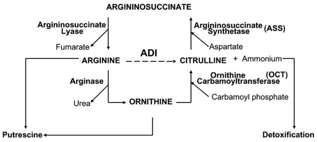
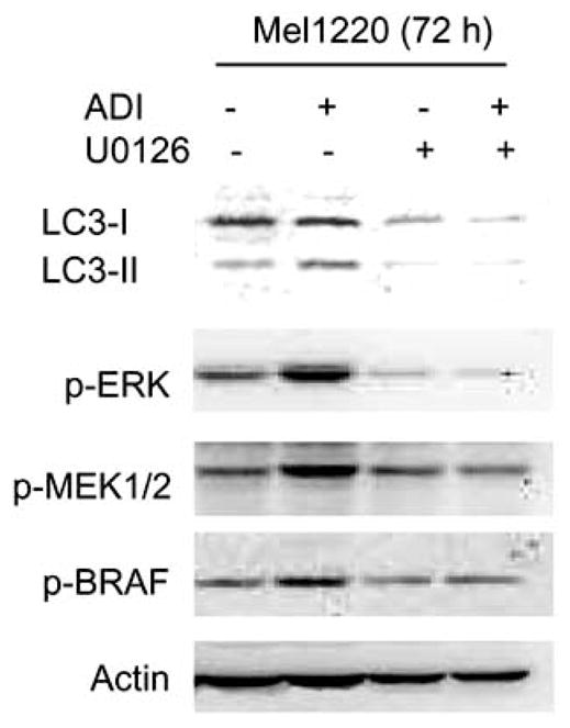
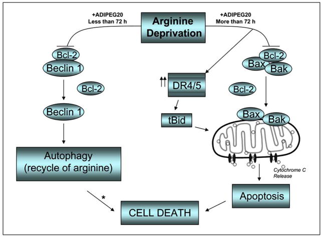
The majority of melanoma cells do not express argininosuccinate synthetase (ASS), and hence cannot synthesize arginine from citrulline. Their growth and proliferation depend on exogenous supply of arginine. Arginine degradation using arginine deiminase (ADI) leads to growth inhibition and eventually cell death while normal cell which express ASS can survive. This notion has been translated into clinical trial. Pegylated ADI (ADI-PEG20) has shown antitumor activity in melanoma. However, the sensitivity to ADI is different among ASS(-) melanoma cells. We have investigated and reviewed the signaling pathways which are affected by arginine deprivation and their consequences which lead to cell death. We have found that arginine deprivation inhibits mTOR signaling but leads to activation of MEK and ERK with no changes in BRAF. These changes most likely lead to autophagy, a possible mechanism to survive by recycling intracellular arginine. However apoptosis does occur which can be both caspase dependent or independent In order to increase the therapeutic efficacy of this form of treatment, one should consider adding other agent(s) which can drive the cells toward apoptosis or inhibit the autophagic process.


Figures

Fig. 1
Fig. 1
Enzymes in urea cycle. The dotted line represents the conversion of arginine to citrulline by ADI, a mycoplasma enzyme.
Fig. 2
Fig. 2
Immunoblot of p-ERK1/2, p-MEK and p-BRAF as well as LC3I-II, a marker for autophagy. The appearance of LC3-II or an increase of LC3-I to II represents autophagic process. Cells were treated with ADI-PEG20 (0.06 ug/ml) alone, U0126 (5 uM) alone or both for 72 h. Note: Treatment with ADI-PEG20 increased MEK and ERK phosphorylation while no changes occurred in BRAF. The addition of MEK inhibitor decreased p-MEK and p-ERK as well as decreased autophagy as detected by the disappearance of LC3-II band.
Fig. 3
Fig. 3
A. Growth inhibitory effect with ADI-PEG20 alone (15%), U0126 alone (25%), and in combination (80%) in a melanoma cell line, Mel1220. B. Apoptosis assay by flow cytometry which detects caspase activity and cell death after treatment with ADI-PEG20 (0.05 ug/ml) and U0126 (5 uM). Note: Treatment with ADI-PEG20 alone or U0126 alone produced only 3–6% of cells which were positive for caspase activity and less than 10% cell death (data not shown), while combination treatment produced 26% of cells which are positive for caspase activity and 30% cell death.
Fig. 4
Fig. 4
A. Autophagy as detected by increased LC3-II by western blot and immunofluorescence in Mel-1220 after treatment with ADI-PEG20 for 72 h, and 5 days at 0.06 ug/ml. B. Apoptosis as detected by increased caspase activity and cell death after 6 days of treatment at 0.1 ug/ml of ADI-PEG20 in Mel1220.
Fig. 5
Fig. 5
Increased cell death after silencing Beclin 1 mRNA. The percentage of cell death increased by 20% upon treatment with 0.1 ug/ml of ADI PEG20 for 3 days.
Fig. 6
Fig. 6
Growth signaling pathway affected by arginine deprivation in ASS(−) cells which leads to autophagy.
Fig. 7
Fig. 7
Possible mechanism of autophagy and apoptosis upon ADI-PEG20 treatment. Treatment with ADI-PEG20 which depletes arginine in the media will first lead to autophagy as a survival mechanism. With longer duration of exposure and higher dose, apoptosis does occur via Bax/Bak activation. * possible link between autophagy and apoptosis (caspase-independent cell death).
Fig. 8
Fig. 8
A. Apoptosis as detected by increased caspase activity and cell death by PI staining in A375 cells. ADI-PEG20 alone at 0.05 ug/ml showed 10% cell death (data not shown), cisplatin alone showed 20% of cells positive for caspase activity and 30% cell death as indicated by PI staining while for the two drug combination the caspase activity increased to 75% and 90% cell death. B. Immunofluorescence of gamma-H2AX (bright dots) which depicts DNA damage. Cisplatin alone showed some increase in gamma-H2AX while the two drug combination showed more cells positive for gamma-H2AX.

References

    1. Copperman R, Morton HE. Proc Soc Exp Biol Med. 1966;123:790–795. - PubMed
    1. Barile MF, Leventhal BG. Nature. 1968;219:750–752. - PubMed
    1. Sugimura K, Fukuda S, Wada Y, Taniai M, Suzuki M, Kimura T, Ohno T, Yamamoto K, Azuma I. Infect Immun. 1990;58:2510–2515. - PMC - PubMed
    1. Sugimura K, Kimura T, Arakawa H, Ohno T, Wada Y, Kimura Y, Saheki T, Azuma I. Leuk Res. 1990;14:931–934. - PubMed
    1. Sugimura K, Ohno T, Fukuda S, Wada Y, Kimura T, Azuma I. Cancer Res. 1990;50:345–349. - PubMed

Publication types

MeSH terms