

Incremental value of contrast myocardial perfusion to detect intermediate versus severe coronary artery stenosis during stress-echocardiography
- PMID: 20459632
- PMCID: PMC2877655
- DOI: 10.1186/1476-7120-8-16
Incremental value of contrast myocardial perfusion to detect intermediate versus severe coronary artery stenosis during stress-echocardiography
Abstract
Background: We aimed to compare the incremental value of contrast myocardial perfusion imaging (MPI) for the detection of intermediate versus severe coronary artery stenosis during dipyridamole-atropine echocardiography (DASE).Wall motion (WM) assessment during stress-echocardiography demonstrates suboptimal sensitivity to detect coronary artery disease (CAD), particularly in patients with isolated intermediate (50%-70%) coronary stenosis.
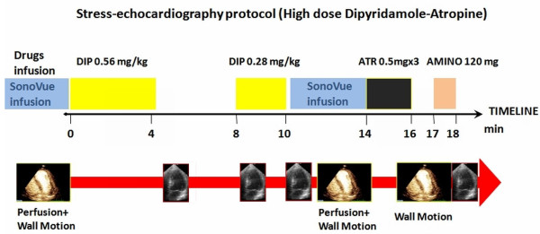
Methods: We performed DASE with MPI in 150 patients with a suspected chest pain syndrome who were given clinical indication to coronary angiography.
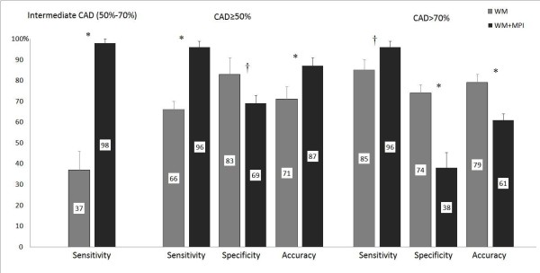
Results and discussion: When CAD was defined as the presence of a >or=50% stenosis, the addition of MPI increased sensitivity (+30%) and decreased specificity (-14%), with a final increase in total diagnostic accuracy (+16%, p < 0.001). The addition of MPI data substantially increased the sensitivity to detect patients with isolated intermediate stenosis from 37% to 98% (p < 0.001); the incremental sensitivity was much lower in patients with severe stenosis, from 85% to 96% (p < 0.05), at the expense of a higher decrease in specificity and a final decrease in total diagnostic accuracy (-18%, p < 0.001).
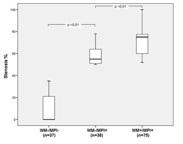
Conclusions: The addition of MPI on top of WM analysis during DASE increases the diagnostic sensitivity to detect obstructive CAD, whatever its definition (>or=50% or > 70% stenosis), but it is mainly driven by the sensitivity increase in the intermediate group (50%-70% stenosis).The total diagnostic accuracy increased only when defining CAD as >or=50% stenosis, since in patients with severe stenosis (> 70%) the decrease in specificity is not counterbalanced by the minor sensitivity increase.
Figures
References
-
- Elhendy A, O'Leary EL, Xie F, McGrain AC, Anderson JR, Porter TR. Comparative accuracy of real-time myocardial contrast perfusion imaging and wall motion analysis during dobutamine stress echocardiography for the diagnosis of coronary artery disease. J Am Coll Cardiol. 2004;44:2185–91. doi: 10.1016/j.jacc.2004.08.059. - DOI - PubMed
-
- Rigo F, Sicari R, Gherardi S, Djordjevic-Dikic A, Cortigiani L, Picano E. Prognostic value of coronary flow reserve in medically treated patients with left anterior descending coronary disease with stenosis 51% to 75% in diameter. Am J Cardiol. 2007;100:1527–31. doi: 10.1016/j.amjcard.2007.06.060. - DOI - PubMed
-
- Nagel E, Lehmkuhl HB, Bocksch W, Klein C, Vogel U, Frantz E, Ellmer A, Dreysse S, Fleck E. Noninvasive diagnosis of ischemia-induced wall motion abnormalities with the use of high-dose dobutamine stress MRI: comparison with dobutamine stress echocardiography. Circulation. 1999;99:763–70. - PubMed
-
- Berger A, Botman KJ, MacCarthy PA, Wijns W, Bartunek J, Heyndrickx GR, Pijls NH, De Bruyne B. Long-term clinical outcome after fractional flow reserve-guided percutaneous coronary intervention in patients with multivessel disease. J Am Coll Cardiol. 2005;46:438–42. doi: 10.1016/j.jacc.2005.04.041. - DOI - PubMed
-
- Wei K, Ragosta M, Thorpe J, Coggins M, Moos S, Kaul S. Noninvasive quantification of coronary blood flow reserve in humans using myocardial contrast echocardiography. Circulation. 2001;103:2560–5. - PubMed
Publication types
MeSH terms
Substances
LinkOut - more resources
Full Text Sources
Miscellaneous
