. 2010 May 7:10:47.
doi: 10.1186/1471-213X-10-47.

Characterization of adipocyte differentiation from human mesenchymal stem cells in bone marrow

Affiliations

Characterization of adipocyte differentiation from human mesenchymal stem cells in bone marrow

Shu-Wen Qian et al. BMC Dev Biol. .

Abstract

Background: Adipocyte hyperplasia is associated with obesity and arises due to adipogenic differentiation of resident multipotent stem cells in the vascular stroma of adipose tissue and remote stem cells of other organs. The mechanistic characterization of adipocyte differentiation has been researched in murine pre-adipocyte models (i.e. 3T3-L1 and 3T3-F442A), revealing that growth-arrest pre-adipocytes undergo mitotic clonal expansion and that regulation of the differentiation process relies on the sequential expression of three key transcription factors (C/EBPbeta, C/EBPalpha and PPARgamma). However, the mechanisms underlying adipocyte differentiation from multipotent stem cells, particularly human mesenchymal stem cells (hBMSCs), remain poorly understood. This study investigated cell cycle regulation and the roles of C/EBPbeta, C/EBPalpha and PPARgamma during adipocyte differentiation from hBMSCs.

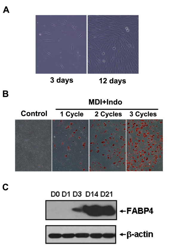
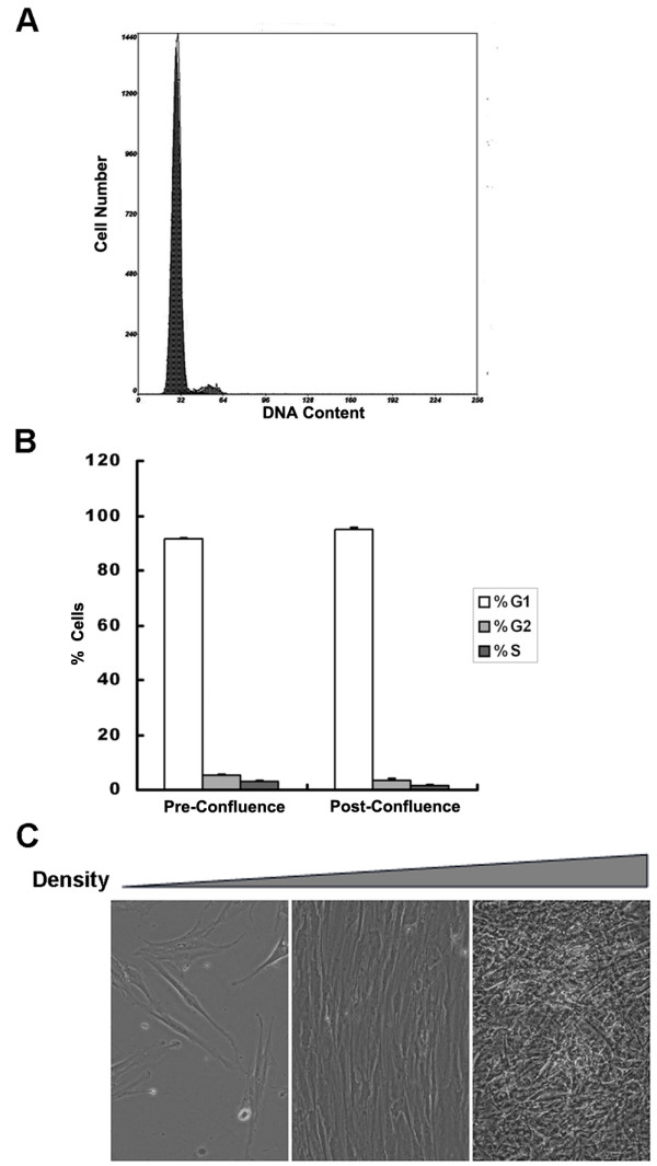
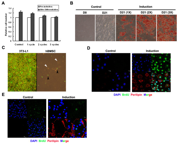
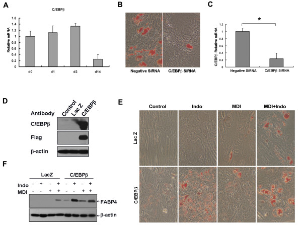
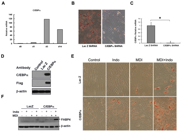
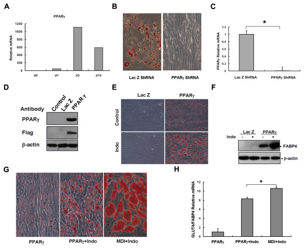
Results: Utilising a BrdU incorporation assay and manual cell counting it was demonstrated that induction of adipocyte differentiation in culture resulted in 3T3-L1 pre-adipocytes but not hBMSCs undergoing mitotic clonal expansion. Knock-down and over-expression assays revealed that C/EBPbeta, C/EBPalpha and PPARgamma were required for adipocyte differentiation from hBMSCs. C/EBPbeta and C/EBPalpha individually induced adipocyte differentiation in the presence of inducers; PPARgamma alone initiated adipocyte differentiation but the cells failed to differentiate fully. Therefore, the roles of these transcription factors during human adipocyte differentiation are different from their respective roles in mouse.

Conclusions: The characteristics of hBMSCs during adipogenic differentiation are different from those of murine cells. These findings could be important in elucidating the mechanisms underlying human obesity further.


Figures

Figure 1
Figure 1
Isolation and adipogenic differentiation of hBMSCs. (A) The morphology of adherent hBMSCs three and 12 days after plating (magnification 100×). (B) HBMSCs of P5 were cultured for one week after confluence and induced to differentiate with MDI+Indo (M: methyl-isobutyl-xanthine; D: dexamethasone; I: insulin; Indo: indomethacin) treatment for one, two or three cycles (1 cycle of treatment: MDI+Indo for three days followed by insulin for one day). The accumulation of cytoplasmic triglyceride was detected by Oil Red O staining on day 21 and visualized under a microscope (magnification 100×). (C) FABP4 expression was examined by Western Blotting at the indicated days after differentiation with repeated MDI+Indo treatment (three times).
Figure 2
Figure 2
Growth characteristic of hBMSCs. (A) Confluent hBMSCs were trypsinized, fixed and stained with PI. DNA content in cells was examined by flow cytometry. (B) Pre-confluent (density ~80%) and post-confluent (one week after cells reach confluence) hBMSCs from three separate experiments at different cell cycle stages revealed by flow cytometry were quantified. (C) Morphology of hBMSCs at different densities (plated at 5000/cm2 and cultured for one day, one week and five weeks).
Figure 3
Figure 3
Cell cycle progression during adipogenic differentiation of hBMSCs. (A) Post-confluence hBMSCs with or without induction (MDI+Indo treatment for one, two or three cycles) were counted and plotted on day 0 and day 21. (B) Cells with parallel treatment in (A) were also stained with oil red O on day 21 and photographed (magnification 100×). (C) 10 μg/ml BrdU was added to 3T3-L1 cells at 18 h after MDI treatment for 30 h, and added to hBMSC at 24 h for 48 h. BrdU incorporation was detected by immunocytochemistry and photographed with both a halogen and mercury lamp switched on (magnification 200×). In 3T3-L1 cells (D) and hBMSCs (E) with or without induction (control), incorporated BrdU (FITC) and fat lipids (TRITC) were shown by confocal microscopy.
Figure 4
Figure 4
C/EBPβ was required for and stimulated adipocyte differentiation from hBMSCs. (A) Relative expression levels of C/EBPβ were determined at the indicated days by real-time PCR. (B) Adipogeic differentiation revealed by oil red O staining with C/EBPβ knock-down by SiRNA. (C) Expression levels of C/EBPβ were determined by real-time PCR (n = 3, *P < 0.05). (D) Over-expression of C/EBPβ in hBMSCs with adenoviral infection (Lac Z as control) was confirmed by Western Blotting. (E) HBMSCs were cultured to confluence and infected with adenovirus at MOI 10 followed by various combination of hormone treatment 4 h later for three days. Cells were stained with oil red O on day eight (magnification 100×). (F) The expression of the adipocyte marker (FABP4) was detected on day four by Western Blotting.
Figure 5
Figure 5
C/EBPα was required for and stimulated adipocyte differentiation from hBMSCs. (A) Relative expression levels of C/EBPα were determined at the indicated days by real-time PCR. (B) Adipocyte differentiation revealed by oil red O staining with C/EBPα knock-down by adenovirus expressing shRNA. (C) C/EBPα knock-down was confirmed by real-time PCR (n = 3, *P < 0.05). (D) C/EBPα over-expression in hBMSCs using adenovirus (Lac Z as control) was shown by Western Blotting. (E) HBMSCs were cultured to confluence and infected with adenovirus at MOI 10 followed by various combinations of hormone treatment 4 h later for three days. Cells were stained with oil red O on day eight (magnification 100×). (F) The expression of the adipocyte marker (FABP4) was detected on day four by Western Blotting.
Figure 6
Figure 6
PPARγ was sufficient to initiate adipocyte differentiation from hBMSCs, but could not induce fully developed adipocytes. (A) Relative expression levels of PPARγ were determined at the indicated days by real-time PCR. (B) Adipogeic differentiation revealed by oil red O staining with PPARγ knock-down by adenovirus expressing shRNA. (C) PPARγ knock-down was verified by real-time PCR (n = 3, *P < 0.05). (D) PPARγ over-expression in hMBSCs with adenovirus (Lac Z as control) was shown by Western Blotting. (E) HBMSCs were cultured to confluence and infected with adenovirus at MOI 10 alone or in combination with indomethacin. Lipid droplets indicated by oil red O staining on day 14 (magnification 100×). (F) The expression of adipocyte marker FABP4 was detected on day six by Western Blotting. (G) Morphology of lipid droplets induced by PPARγ expression and hormone treatment (magnification 200×). (H) GLUT4 expression normalized by FABP4 was quantified by real-time PCR in cells treated with PPARγ adenovirus or hormone (n = 3, *P < 0.05).

Similar articles

Cited by

References

    1. Hirsch J, Batchelor B. Adipose tissue cellularity in human obesity. Clin Endocrinol Metab. 1976;5:299–311. doi: 10.1016/S0300-595X(76)80023-0. - DOI - PubMed
    1. Shepherd PR, Gnudi L, Tozzo E, Yang H, Leach F, Kahn BB. Adipose cell hyperplasia and enhanced glucose disposal in transgenic mice overexpressing GLUT4 selectively in adipose tissue. J Biol Chem. 1993;268:22243–22246. - PubMed
    1. Yu ZK, Wright JT, Hausman GJ. Preadipocyte recruitment in stromal vascular cultures after depletion of committed preadipocytes by immunocytotoxicity. Obesity Res. 1997;5:9–15. - PubMed
    1. Crisan M, Yap S, Casteilla L, Chen CW, Corselli M, Park TS, Andriolo G, Sun B, Zheng B, Zhang L, Norotte C, Teng PN, Traas J, Schugar R, Deasy BM, Badylak S, Buhring HJ, Giacobino JP, Lazzari L, Huard J, Pe'ault B. A Perivascular Origin for Mesenchymal Stem Cells in Multiple Human Organs. Cell Stem Cell. 2008;3:301–313. doi: 10.1016/j.stem.2008.07.003. - DOI - PubMed
    1. Crossno JT Jr, Majka SM, Grazia T, Gill RG, Klemm DJ. Rosiglitazone promotes development of a novel adipocyte population from bone marrow-derived circulating progenitor cells. J Clin Invest. 2006;116:3220–8. doi: 10.1172/JCI28510. - DOI - PMC - PubMed

Publication types

Substances

LinkOut - more resources