

The expression and significance of IDH1 and p53 in osteosarcoma
- PMID: 20459648
- PMCID: PMC2873426
- DOI: 10.1186/1756-9966-29-43
The expression and significance of IDH1 and p53 in osteosarcoma
Abstract
Background: To detect the expression of isocitrate dehydrogenase 1 (IDH1) and transformation-related protein 53 (p53) in osteosarcoma and analyze the correlation between them and the clinico-pathological features.
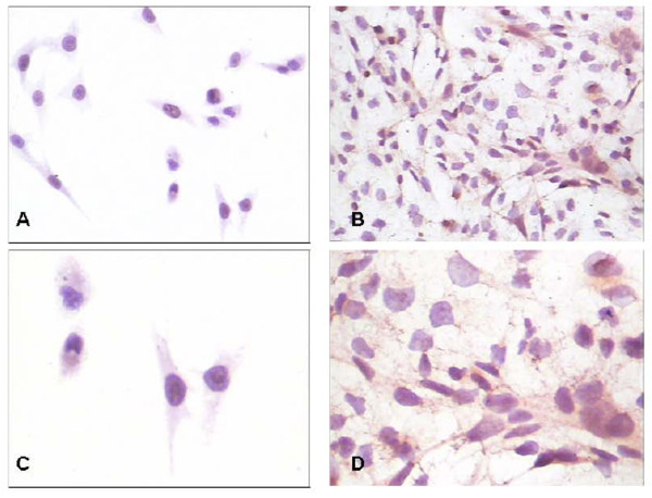
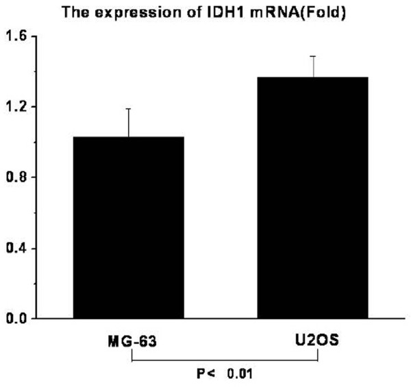
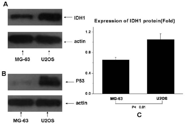
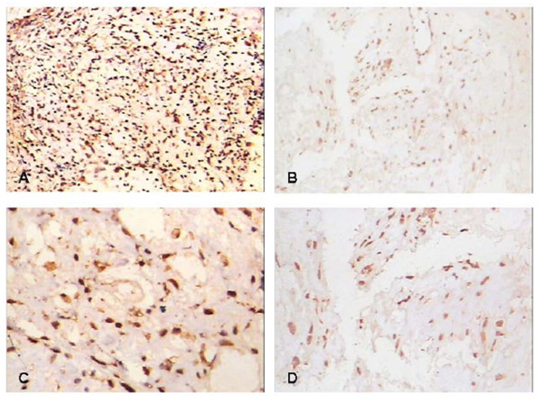
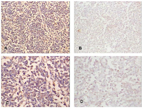
Methods: The expressions of IDH1 and p53 were detected in human osteosarcoma cell lines (MG-63 and U2OS) by immunocytochemistry, Real-time PCR and Western Blotting. The expressions of IDH1 and p53 in formalin-fixed paraffin-embedded tissue sections from 44 osteosarcoma patients were determined by immunohistochemistry, and the correlation between them and clinicopagthological features were analyzed. None of these patients received chemotherapy prior to surgery.
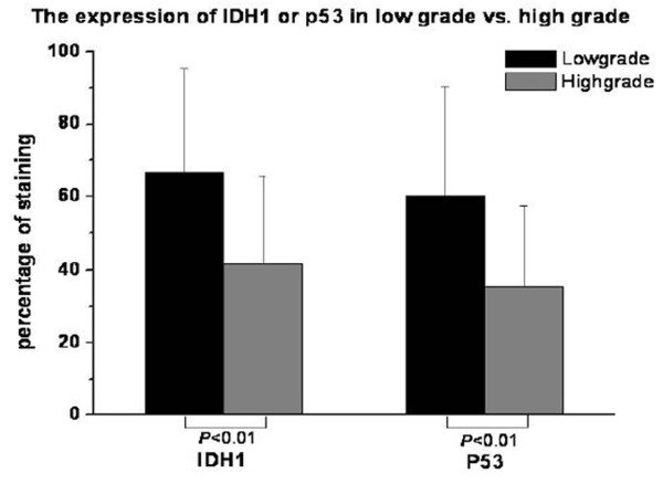
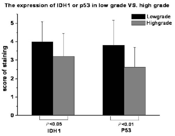
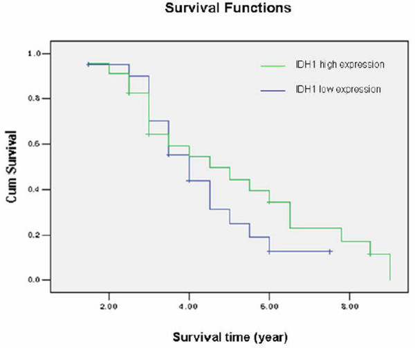
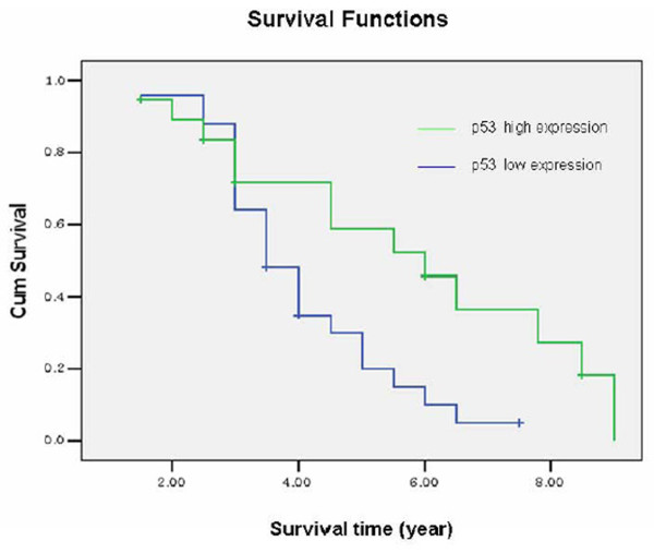
Results: IDH1 is detected in osteosarcoma cell lines and biopsies. IDH1 expresses higher in U2OS cells with wild type p53 than in MG-63 cells with mutation p53. IDH1 correlates with histological Rosen grade and metastasis negatively. P53 correlates with histological Rosen grade, metastasis and overall survival in clinical osteosarcoma biopsies. Osteosarcoma patients with High IDH1 expression have a very high p53 expression.
Conclusion: IDH1 may correlate with p53 and be a candidate biomarker for osteosarcoma correlate with histological Rosen grade and metastasis.
Figures
Similar articles
-
Expressions of p53, c-MYC, BCL-2 and apoptotic index in human osteosarcoma and their correlations with prognosis of patients.Cancer Epidemiol. 2012 Apr;36(2):212-6. doi: 10.1016/j.canep.2011.08.002. Epub 2011 Sep 3. Cancer Epidemiol. 2012. PMID: 21890444
-
Up-regulated isocitrate dehydrogenase 1 suppresses proliferation, migration and invasion in osteosarcoma: in vitro and in vivo.Cancer Lett. 2014 Apr 28;346(1):114-21. doi: 10.1016/j.canlet.2013.12.020. Epub 2013 Dec 22. Cancer Lett. 2014. PMID: 24368190 Free PMC article.
-
AHA1 upregulates IDH1 and metabolic activity to promote growth and metastasis and predicts prognosis in osteosarcoma.Signal Transduct Target Ther. 2021 Jan 20;6(1):25. doi: 10.1038/s41392-020-00387-1. Signal Transduct Target Ther. 2021. PMID: 33468990 Free PMC article.
-
Meta-analysis of clinical significance of p53 protein expression in patients with osteosarcoma.Future Oncol. 2017 Sep;13(21):1883-1891. doi: 10.2217/fon-2017-0180. Epub 2017 Aug 2. Future Oncol. 2017. PMID: 28766969
-
Expression of mutated isocitrate dehydrogenase-1 in gliomas is associated with p53 and EGFR expression.Folia Neuropathol. 2011;49(2):88-93. Folia Neuropathol. 2011. PMID: 21845536
Cited by
-
Osteosarcoma of jaws.J Oral Maxillofac Pathol. 2012 May;16(2):233-8. doi: 10.4103/0973-029X.99075. J Oral Maxillofac Pathol. 2012. PMID: 22923896 Free PMC article.
-
Metformin sensitizes endometrial cancer cells to chemotherapy through IDH1-induced Nrf2 expression via an epigenetic mechanism.Oncogene. 2018 Oct;37(42):5666-5681. doi: 10.1038/s41388-018-0360-7. Epub 2018 Jun 19. Oncogene. 2018. PMID: 29921847
-
The regulatory mechanisms and inhibitors of isocitrate dehydrogenase 1 in cancer.Acta Pharm Sin B. 2023 Apr;13(4):1438-1466. doi: 10.1016/j.apsb.2022.12.019. Epub 2023 Feb 2. Acta Pharm Sin B. 2023. PMID: 37139412 Free PMC article. Review.
-
Prognostic significance of p53 expression in malignant bone tumors: a meta-analysis.Tumour Biol. 2013 Apr;34(2):1037-43. doi: 10.1007/s13277-012-0643-5. Epub 2013 Jan 23. Tumour Biol. 2013. PMID: 23341181
-
Prognostic value of p53 alterations in human osteosarcoma: a meta analysis.Int J Clin Exp Pathol. 2014 Sep 15;7(10):6725-33. eCollection 2014. Int J Clin Exp Pathol. 2014. PMID: 25400752 Free PMC article. Review.
References
-
- Kager L, Zoubek A, Pötschger U, Kastner U, Flege S, Kempf-Bielack B, Branscheid D, Kotz R, Salzer-Kuntschik M, Winkelmann W, Jundt G, Kabisch H, Reichardt P, Jürgens H, Gadner H, Bielack SS. Primary metastatic osteosarcoma: presentation and outcome of patients treated on neoadjuvant Cooperative Osteosarcoma Study Group protocols. J Clin Oncol. 2003;21:2011–2018. doi: 10.1200/JCO.2003.08.132. - DOI - PubMed
-
- Overholtzer M, Rao PH, Favis R, Lu XY, Elowitz MB, Barany F, Ladanyi M, Gorlick R, Levine AJ. The presence of p53 mutations in human osteosarcomas correlates with high levels of genomic instability. Proceedings of the National Academy of Sciences of the United States of America. 2003;100:11547–11552. doi: 10.1073/pnas.1934852100. - DOI - PMC - PubMed
Publication types
MeSH terms
Substances
LinkOut - more resources
Full Text Sources
Medical
Research Materials
Miscellaneous
