. 2010 May 8:9:102.
doi: 10.1186/1476-4598-9-102.

Cadmium induces Wnt signaling to upregulate proliferation and survival genes in sub-confluent kidney proximal tubule cells

Affiliations

Cadmium induces Wnt signaling to upregulate proliferation and survival genes in sub-confluent kidney proximal tubule cells

Prabir K Chakraborty et al. Mol Cancer. .

Abstract

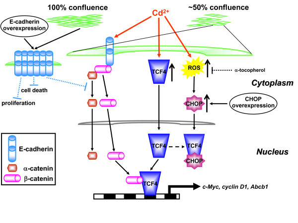
Background: The class 1 carcinogen cadmium (Cd2+) disrupts the E-cadherin/beta-catenin complex of epithelial adherens junctions (AJs) and causes renal cancer. Deregulation of E-cadherin adhesion and changes in Wnt/beta-catenin signaling are known to contribute to carcinogenesis.

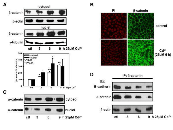
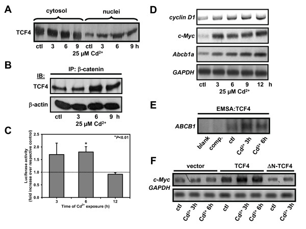
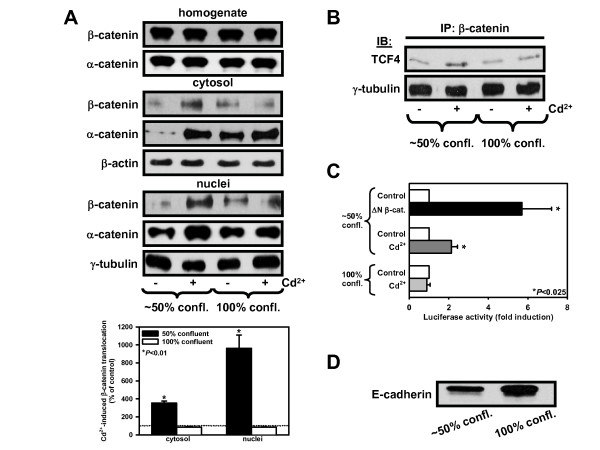
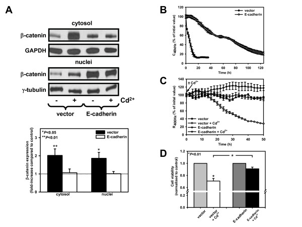
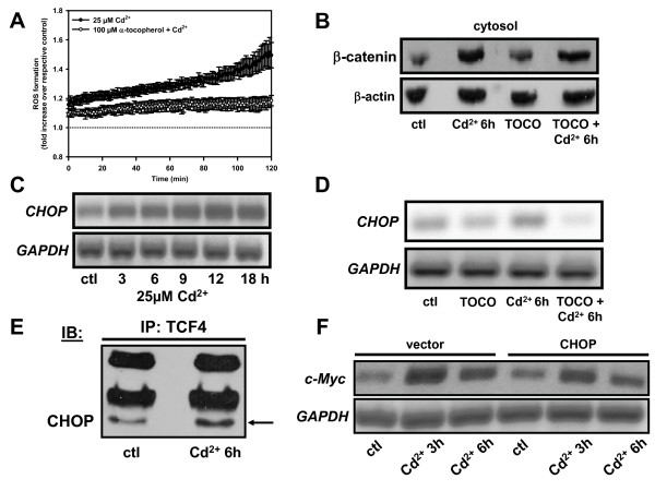
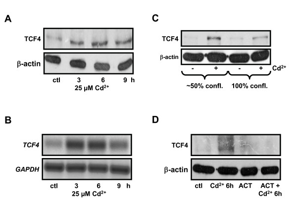
Results: We investigated Wnt signaling after Cd2+-induced E-cadherin disruption in sub-confluent cultured kidney proximal tubule cells (PTC). Cd2+ (25 microM, 3-9 h) caused nuclear translocation of beta-catenin and triggered a Wnt response measured by TOPflash reporter assays. Cd2+ reduced the interaction of beta-catenin with AJ components (E-cadherin, alpha-catenin) and increased binding to the transcription factor TCF4 of the Wnt pathway, which was upregulated and translocated to the nucleus. While Wnt target genes (c-Myc, cyclin D1 and ABCB1) were up-regulated by Cd2+, electromobility shift assays showed increased TCF4 binding to cyclin D1 and ABCB1 promoter sequences with Cd2+. Overexpression of wild-type and mutant TCF4 confirmed Cd2+-induced Wnt signaling. Wnt signaling elicited by Cd2+ was not observed in confluent non-proliferating cells, which showed increased E-cadherin expression. Overexpression of E-cadherin reduced Wnt signaling, PTC proliferation and Cd2+ toxicity. Cd2+ also induced reactive oxygen species dependent expression of the pro-apoptotic ER stress marker and Wnt suppressor CHOP/GADD153 which, however, did not abolish Wnt response and cell viability.

Conclusions: Cd2+ induces Wnt signaling in PTC. Hence, Cd2+ may facilitate carcinogenesis of PTC by promoting Wnt pathway-mediated proliferation and survival of pre-neoplastic cells.


Figures

Figure 1
Figure 1
Cd2+ disrupts the adherens junction complex and redistributes β-catenin from the periphery to cytosol and nuclei of kidney PTC. (A) Time dependent increase of β-catenin protein distribution in cytoplasmic and nuclear fractions of WKPT-0293 Cl.2 cells without (ctl) or with Cd2+. Cytosolic and nuclear fractions were immunoblotted with β-catenin antiserum. For loading controls, the same membranes were reprobed with antibodies to β-actin and γ-tubulin for cytoplasmic and nuclear fractions, respectively. β-catenin signals were normalized to loading markers and compared to controls, which were set to 100%. Means ± SEM of 3 experiments are shown. One-way ANOVA was used assuming equality of variance with Levene's test and Tukey post hoc test for pair-wise comparison between control and Cd2+ exposed cells. (B) Immunofluorescence staining patterns of β-catenin in WKPT-0293 Cl.2 cells ± Cd2+. Nuclei were stained with propidium iodide (PI). Note the peripheral β-catenin labeling in control cells whereas in Cd2+ exposed cells β-catenin is found diffusely distributed in cytosol and nuclei. Bars = 20 μm. (C) Distribution of α-catenin in cytoplasmic and nuclear fractions increases with Cd2+ exposure time, as shown by immunoblotting. (D) Cd2+ reduces association of β-catenin with E-cadherin and α-catenin as a function of time. Cell lysates were immunoprecipitated (IP) with β-catenin antiserum and immunoblotted (IB) with E-cadherin or α-catenin antisera.
Figure 2
Figure 2
Cd2+ triggers nuclear translocation of TCF4 and activates TCF4/β-catenin mediated transcription of cell proliferation and survival genes in kidney PTC. (A) Distribution of TCF4 protein in cytoplasmic and nuclear fractions of WKPT-0293 Cl.2 cells ± Cd2+ was determined by immunoblotting. (B) Increased association of immunoprecipitated β-catenin with TCF4 by Cd2+ in WKPT-0293 Cl.2 cells. (C) Cd2+ induces TCF transcriptional activity. Luciferase activity of TOPflash or FOPflash transfected cells treated with 25 μM Cd2+ for 3-12 h. Data were corrected for protein and normalized to controls at each time point. Means ± SEM (n = 3-8) are shown. Student's unpaired t-test compares Cd2+ treated cells to respective controls. (D) Effect of Cd2+ exposure on mRNA of Wnt pathway target genes. GAPDH was used as housekeeping gene. (E) EMSA of TCF4 binding to the ABCB1 promoter region. Extracts from controls (ctl), Cd2+ (25 μM) treated cells, cells exposed to 3 h Cd2+ followed by incubation with a 200-fold excess of competing unlabeled oligonucleotides (comp.), and lysate-free sample (blank) were loaded. Binding of oligonucleotides to TCF4 was enhanced upon Cd2+ exposure. (F) Effect of TCF4 overexpression on Cd2+ induced c-Myc mRNA expression. WKPT-0293 Cl.2 cells transiently transfected with full length human TCF4 (TCF4), human TCF4 lacking the interaction domain for β-catenin (ΔN-TCF4), or empty vector were treated with ± 25 μM Cd2+. TCF4 overexpression enhanced basal and Cd2+-induced c-Myc expression, whereas ΔN-TCF4 had no effect.
Figure 3
Figure 3
Cd2+-induced nuclear translocation of β-catenin and Wnt signaling are abolished in confluent kidney PTC, which show increased E-cadherin expression. (A) Immunoblots with β-catenin or α-catenin antisera in ~50% and 100% confluent WKPT-0293 Cl.2 cells (25 μM Cd2+, 6 h). For statistical analysis of immunoblots β-catenin signals were normalized to the loading markers and compared to controls, which were set to 100%. Means ± SEM (n = 3) are shown. Student's unpaired t-test compares Cd2+ treated cells to respective controls. (B) Cd2+-induced TCF transcriptional activity depends on the confluence of kidney PTC. WKPT-0293 Cl.2 cells transfected with TOPflash, FOPflash or ΔN-β-catenin were treated with 25 μM Cd2+ for 6 h. Data presented are means ± SEM of 4-5 experiments. Student's unpaired t-test compares Cd2+ treated or ΔN-β-catenin-transfected cells to respective controls. (C) To determine association of TCF4 with β-catenin in ~50% and 100% confluent cells, nuclear fractions were immunoprecipitated with an antibody against β-catenin and immunoblotted for TCF4. (D) Expression of E-cadherin in whole cell lysates of ~50% and 100% confluent cells by immunoblotting.
Figure 4
Figure 4
E-cadherin overexpression in kidney PTC abolishes β-catenin redistribution, decreases proliferation and protects against Cd2+-induced epithelial barrier disruption and cytotoxicity. (A)β-catenin immunoblots of WKPT-0293 Cl.2 cells transiently transfected with full length E-cadherin expression plasmid (E-cadherin) or empty vector (vector) and incubated for 6 h ± 25 μM Cd2+. β-catenin signals were normalized to loading markers and the ratio of β-catenin in Cd2+ and respective control samples was determined. Means ± SEM (n = 7) are shown. Student's unpaired t-test compares Cd2+ treated cells to respective controls. (B) Vector or E-cadherin PTC were reseeded in SCM in ECIS 8WE10 arrays. C40 kHz reflects cell attachment, spreading and proliferation (see Methods). Initial values of the cell-free electrode were set to 100% (vector, 63.5 ± 1.9 nF; E-cadherin, 65.2 ± 3.3 nF; means ± SEM of 3 experiments). Data show means ± SEM of 3 experiments. (C) At confluence PTC were exposed to SFM ± 20 μM Cd2+. Values of the confluent cell-covered electrode were set to 100% (vector, 3670 ± 83 ohm; E-cadherin, 2994 ± 300 ohm; means ± SEM of 3 different experiments). A drop in R400 Hz indicates disruption of epithelial barrier and cell detachment. (D) Vector or E-cadherin cells were exposed to 25 μM Cd2+ for 6 h and cell viability was determined by MTT assay. Graph depicts means ± SEM of 9-10 experiments. Student's unpaired t-test compares Cd2+ treated cells to respective controls as well as Cd2+-exposed vector to Cd2+-exposed E-cadherin cells.
Figure 5
Figure 5
Cd2+-induced reactive oxygen species (ROS) formation in kidney PTC induces up-regulation of CHOP, but has no effect on Wnt signaling. (A) When used, the ROS scavenger α-tocopherol was pre-incubated for 1 h. Cells were loaded with 20 μM carboxy-H2DCFDA. Cell suspensions were incubated with Cd2+ ± α-tocopherol and fluorescent signals were measured immediately at 500/535 nm. Graph shows means ± SEM of 4-7 experiments. (B) β-catenin immunoblot of WKPT-0293 Cl.2 cells ± α-tocopherol (TOCO) (100 μM) ± 25 μM Cd2+ for 6 h. (C) Expression of CHOP mRNA in WKPT-0293 Cl.2 cells without (ctl) or with 25 μM Cd2+ by RT-PCR. (D) Prevention of Cd2+-mediated increase in CHOP mRNA expression by α-tocopherol. (E) Association of CHOP with TCF4 is increased in WKPT-0293 Cl.2 cells incubated with 25 μM Cd2+ for 6 h when compared to controls (ctl). (F) Effect of CHOP overexpression on Cd2+-induced expression of c-Myc by RT-PCR. PTC transiently transfected with full length human CHOP (CHOP) or empty vector (vector) were exposed to 25 μM Cd2+.
Figure 6
Figure 6
Cd2+ triggers TCF4 up-regulation in kidney PTC. (A) TCF4 protein expression was increased by Cd2+ in whole cell lysates from PTC. (B) RT-PCR of mRNA was performed with primers specific for rat TCF4 in WKPT-0293 Cl.2 cells without (ctl) or with Cd2+. Cd2+ increased TCF4 gene expression. (C) TCF4 expression in whole cell lysates of ~50% confluent and 100% confluent cells ± Cd2+ (25 μM for 6 h) by immunoblotting confirmed TCF4 up-regulation induced by Cd2+ in subconfluent cells. (D) Actinomycin D abolished Cd2+-induced TCF4 up-regulation. PTC were treated with or without Cd2+ (25 μM) ± actinomycin D (ACT) (10 μg/ml, 1 h pre-incubation), a transcriptional inhibitor. Expression of TCF4 protein was determined in whole cell lysates of WKPT-0293 Cl.2 cells by immunoblotting.
Figure 7
Figure 7
Model for the effects of Cd2+ on the adherens junction complex and Wnt signaling in WKPT-0293 Cl.2 kidney PTC. For further details, see discussion.

Similar articles

Cited by

References

    1. Angers S, Moon RT. Proximal events in Wnt signal transduction. Nat Rev Mol Cell Biol. 2009;10:468–477. - PubMed
    1. Clevers H. Wnt/beta-catenin signaling in development and disease. Cell. 2006;127:469–480. doi: 10.1016/j.cell.2006.10.018. - DOI - PubMed
    1. Shapiro L, Weis WI. Structure and biochemistry of cadherins and catenins. Cold Spring Harb Perspect Biol. 2009;1 doi: 10.1101/cshperspect.a003053. - PMC - PubMed
    1. Miyoshi J, Takai Y. Molecular perspective on tight-junction assembly and epithelial polarity. Adv Drug Deliv Rev. 2005;57:815–855. doi: 10.1016/j.addr.2005.01.008. - DOI - PubMed
    1. Danilkovitch-Miagkova A, Miagkov A, Skeel A, Nakaigawa N, Zbar B, Leonard EJ. Oncogenic mutants of RON and MET receptor tyrosine kinases cause activation of the beta-catenin pathway. Mol Cell Biol. 2001;21:5857–5868. doi: 10.1128/MCB.21.17.5857-5868.2001. - DOI - PMC - PubMed

Publication types

MeSH terms