. 2010 May 10:8:57.
doi: 10.1186/1741-7007-8-57.

Controlled spatial and conformational display of immobilised bone morphogenetic protein-2 and osteopontin signalling motifs regulates osteoblast adhesion and differentiation in vitro

Affiliations

Controlled spatial and conformational display of immobilised bone morphogenetic protein-2 and osteopontin signalling motifs regulates osteoblast adhesion and differentiation in vitro

Elizabeth A Mitchell et al. BMC Biol. .

Abstract

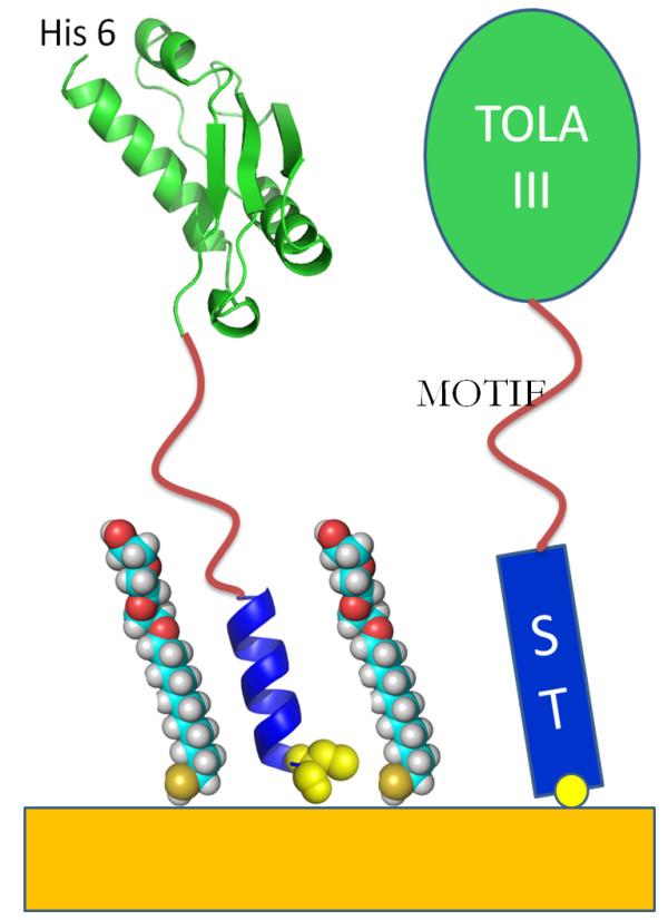
Background: The interfacial molecular mechanisms that regulate mammalian cell growth and differentiation have important implications for biotechnology (production of cells and cell products) and medicine (tissue engineering, prosthetic implants, cancer and developmental biology). We demonstrate here that engineered protein motifs can be robustly displayed to mammalian cells in vitro in a highly controlled manner using a soluble protein scaffold designed to self assemble on a gold surface.

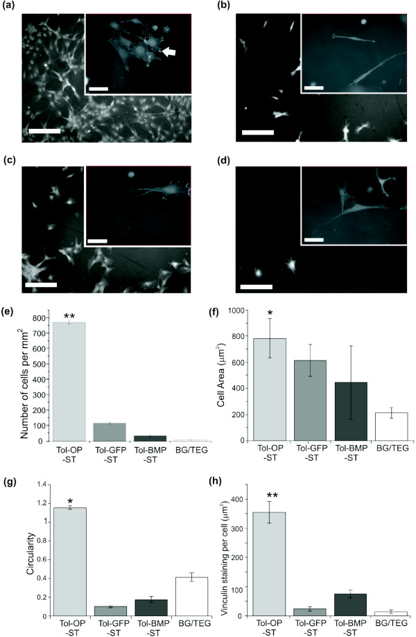
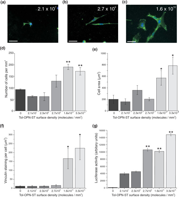
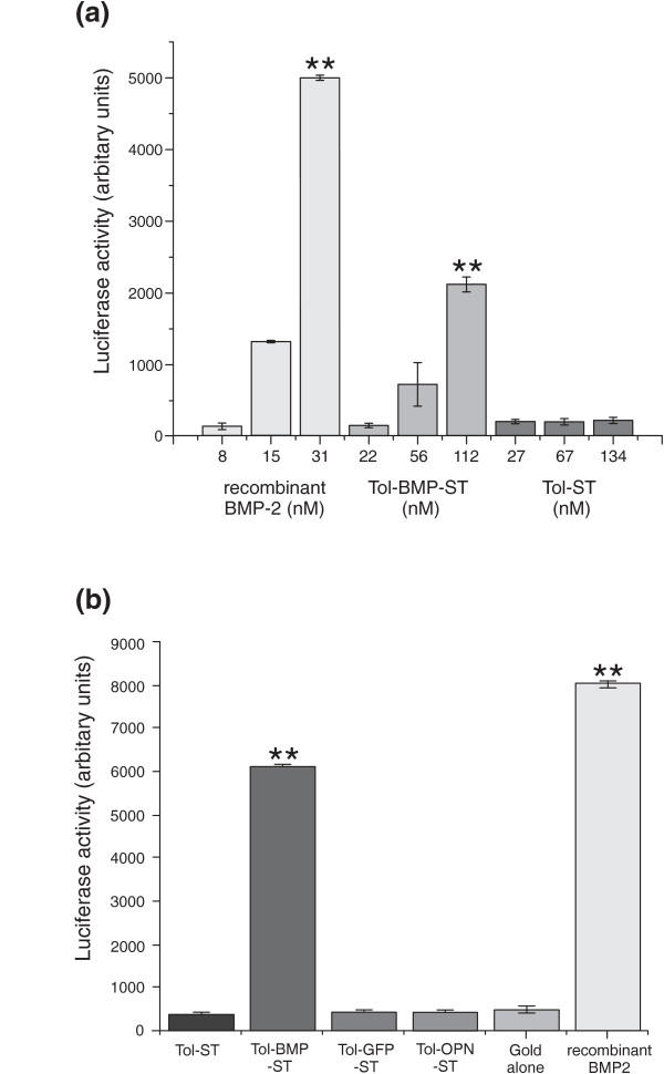
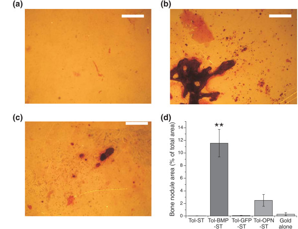
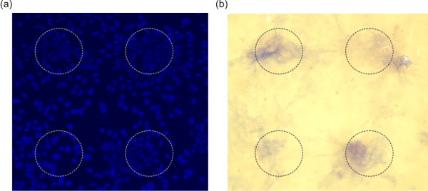
Results: A protein was engineered to contain a C-terminal cysteine that would allow chemisorption to gold, followed by 12 amino acids that form a water soluble coil that could switch to a hydrophobic helix in the presence of alkane thiols. Bioactive motifs from either bone morphogenetic protein-2 or osteopontin were added to this scaffold protein and when assembled on a gold surface assessed for their ability to influence cell function. Data demonstrate that osteoblast adhesion and short-term responsiveness to bone morphogenetic protein-2 is dependent on the surface density of a cell adhesive motif derived from osteopontin. Furthermore an immobilised cell interaction motif from bone morphogenetic protein supported bone formation in vitro over 28 days (in the complete absence of other osteogenic supplements). In addition, two-dimensional patterning of this ligand using a soft lithography approach resulted in the spatial control of osteogenesis.

Conclusion: These data describe an approach that allows the influence of immobilised protein ligands on cell behaviour to be dissected at the molecular level. This approach presents a durable surface that allows both short (hours or days) and long term (weeks) effects on cell activity to be assessed. This widely applicable approach can provide mechanistic insight into the contribution of immobilised ligands in the control of cell activity.


Figures

Figure 1
Figure 1
The design of the Tol switch tag system, a molecular image (left) and a schematic (right). The protein starts at the N-terminus with a 6 His tag for easy purification followed by a FLAG-tag sequence to aid detection. The TolAIII domain which comes next provides solubility and drives high levels of expression for fusion proteins attached to its C terminus [10]. The fusion sequence is inserted between the TolA and the switch tag, which is found at the C terminus, by use of a multiple cloning site in the plasmid [8]. The TolA domain can be removed by specific proteolytic cleavage but in most cases it is left in place since it stabilises the fusion construct. The switch tag is shown here as a helix whose hydrophobic properties allow co assembly with alkane thiol SAM, in solution it is a water soluble random coil.
Figure 2
Figure 2
Immobilised Tol scaffold containing the osteopontin motif, SVVYGLR, specifically controls cell adhesion and cell morphology. Cells were plated onto surfaces displaying scaffold proteins in the absence of serum supplements and assessed after 24 hours. Immunolocalization of vinculin and counterstaining with DAPI allowed identification of focal adhesion complexes (white arrow) in cells on (a) Tol-OPN-ST; (b) Tol-GFP-ST; (c) Tol-BMP-ST; (d) TEG. Size bars = 200 μm and 50 μm in the inset images. Image analysis demonstrated significant differences in (e) the number of cells adhered to Tol-OPN-ST compared to all other surfaces; (f) the area of cells on Tol-OPN-ST compared to TEG; (g) the circularity of cells on Tol-OPN-ST compared to the other surfaces and (h) abundance of vinculin positivity in cells on Tol-OPN-ST compared to other surfaces. Data representative of three independent experiments; results are mean ± SEM; *P < 0.05, ** P < 0.01. Significance was determined by ANOVA with Fisher post test.
Figure 3
Figure 3
Tol-OP-ST surface density influences cell morphology and response to BMP2. Gold coverslips were incubated with different concentrations of Tol-OP-ST and based on SPR analysis surface densities calculated (molecules/mm2). (a, b, c) Cells were plated onto the surfaces, as indicated, under serum free conditions and vinculin immunolocalized 24 hours later. Size bars = 50 μm. (d) DAPI was used to visualize nuclear DNA and images quantified to show number of adhered cells. Numbers of cells on 1.6 and 3.3 × 10¹⁰ molecules/mm2 were significantly different to all other surfaces. (e) Assessment of cell area revealed that cells on 1.6 and 3.3 × 10¹⁰ molecules/mm2 were significantly different to all other surfaces. Furthermore (f) levels of vinculin positivity were significantly higher in cells on 1.6 and 3.3 × 10¹⁰ molecules/mm2 in comparison to lower densities. (g) Cells transfected with a SMAD-reporter construct were plated on the surfaces, treated with 100 ng/ml recombinant BMP-2 and levels of luciferase assessed and normalised to cell number. Data representative of three independent experiments; results are mean ± SEM; * P < 0.05, ** P < 0.01. Significance was determined by ANOVA with Fisher post test.
Figure 4
Figure 4
Tol scaffold containing the BMP2 knuckle peptide, KIPKASSVPTELSAISTLYL, activates Smad dependent signalling both when added exogenously to adherent cells and when immobilised on a surface. (a) Cells were cultured in multiwell plates, transfected with a SMAD-reporter construct and then treated with recombinant BMP2, exogenous Tol-BMP-ST or Tol-ST. Levels of luciferase were then assessed after eight hours. Both recombinant BMP2 (31 nM) and Tol-BMP-ST (112 nM) induced significant levels of luciferase activity compared to Tol alone. (b) Cells already transfected with the SMAD-reporter construct were plated onto gold coverslips displaying the scaffold proteins at the concentrations outlined in Table 1 or TEG alone. Luciferase activity was measured 12 hours later and contrasted with the same cells grown on tissue culture plastic and treated with soluble recombinant BMP-2 (100 ng/ml). Significant levels of SMAD signalling were only observed on the Tol-BMP-ST surface. Data representative of three independent experiments; results are mean ± SEM; ** P < 0.01. Significance was determined by ANOVA with Tukey post test.
Figure 5
Figure 5
Long term growth of calvarial-derived cells on scaffold proteins. After four weeks under routine culture conditions and in the absence of exogeneous osteogenic factors mineralized matrix was visualized with Alizarin red staining. There was little evidence of mineralised matrix on (a) Tol alone, whilst abundant deposition was observed on (b) Tol-BMP-ST and a small amount on (c) Tol-OP-ST surfaces. Size bar = 400 μm. (d) Image analysis shows significant evidence of mineralised matrix deposition only on Tol-BMP surfaces. Data representative of three independent experiments; results are mean ± SEM; ** P < 0.01. Significance was determined by ANOVA with Tukey post test.
Figure 6
Figure 6
Micro-patterned Tol-BMP-ST supported spatially restricted differentiation of calvarial-derived cells. Tol-BMP-St was patterned onto gold surfaces using a soft lithography approach to create 150 μm diameter circles of scaffold protein. Cells were cultured on these surfaces for 14 days. (a) Cell distribution was assessed by DAPI staining for nuclear DNA and (b) cell differentiation was investigated by alkaline phosphatase staining (blue).

Comment in

Similar articles

Cited by

References

    1. Daley WP, Peters SB, Larsen M. Extracellular matrix dynamics in development and regenerative medicine. J Cell Sci. 2008;121:255–264. doi: 10.1242/jcs.006064. - DOI - PubMed
    1. Pierschbacher MD, Ruoslahti E. Variants of the cell recognition site of fibronectin that retain attachment-promoting activity. Proc Natl Acad Sci USA. 1984;81:5985–5988. doi: 10.1073/pnas.81.19.5985. - DOI - PMC - PubMed
    1. Humphries MJ, Obara M, Olden K, Yamada KM. Role of fibronectin in adhesion, migration, and metastasis. Cancer Invest. 1989;7:373–393. doi: 10.3109/07357908909039866. - DOI - PubMed
    1. Garamszegi N, Garamszegi SP, Shehadeh LA, Scully SP. Extracellular matrix-induced gene expression in human breast cancer cells. Mol Cancer Res. 2009;7:319–329. doi: 10.1158/1541-7786.MCR-08-0227. - DOI - PMC - PubMed
    1. Sahni A, Sporn LA, Francis CW. Potentiation of endothelial cell proliferation by fibrin(ogen)-bound fibroblast growth factor-2. J Biol Chem. 1999;274:14936–14941. doi: 10.1074/jbc.274.21.14936. - DOI - PubMed

Publication types