. 2010 May 10:11:35.
doi: 10.1186/1471-2199-11-35.

Analysis of illegitimate genomic integration mediated by zinc-finger nucleases: implications for specificity of targeted gene correction

Affiliations

Analysis of illegitimate genomic integration mediated by zinc-finger nucleases: implications for specificity of targeted gene correction

Petter A Olsen et al. BMC Mol Biol. .

Abstract

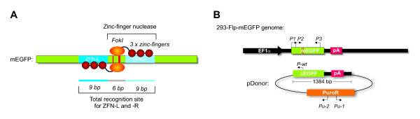
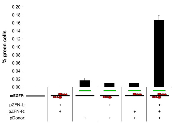
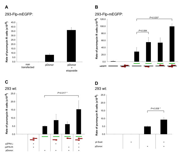
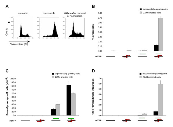
Background: Formation of site specific genomic double strand breaks (DSBs), induced by the expression of a pair of engineered zinc-finger nucleases (ZFNs), dramatically increases the rates of homologous recombination (HR) between a specific genomic target and a donor plasmid. However, for the safe use of ZFN induced HR in practical applications, possible adverse effects of the technology such as cytotoxicity and genotoxicity need to be well understood. In this work, off-target activity of a pair of ZFNs has been examined by measuring the ratio between HR and illegitimate genomic integration in cells that are growing exponentially, and in cells that have been arrested in the G2/M phase.

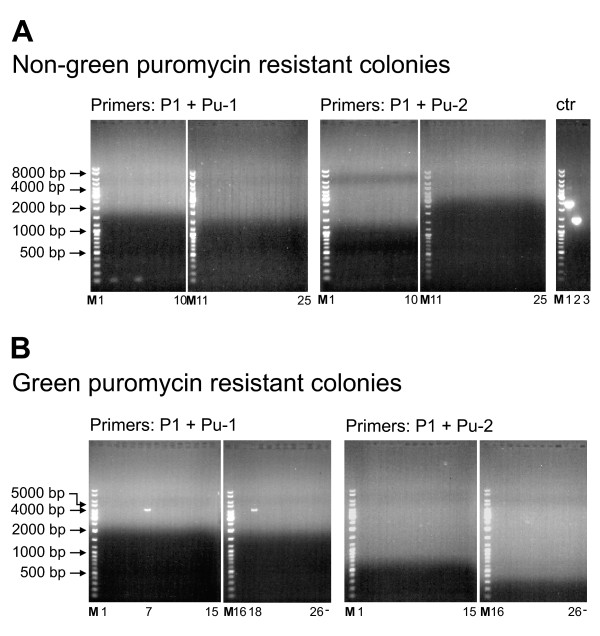
Results: A reporter cell line that contained consensus ZFN binding sites in an enhanced green fluorescent protein (EGFP) reporter gene was used to measure ratios between HR and non-homologous integration of a plasmid template. Both in human cells (HEK 293) containing the consensus ZFN binding sites and in cells lacking the ZFN binding sites, a 3.5 fold increase in the level of illegitimate integration was observed upon ZFN expression. Since the reporter gene containing the consensus ZFN target sites was found to be intact in cells where illegitimate integration had occurred, increased rates of illegitimate integration most likely resulted from the formation of off-target genomic DSBs. Additionally, in a fraction of the ZFN treated cells the co-occurrence of both specific HR and illegitimate integration was observed. As a mean to minimize unspecific effects, cell cycle manipulation of the target cells by induction of a transient G2/M cell cycle arrest was shown to stimulate the activity of HR while having little effect on the levels of illegitimate integration, thus resulting in a nearly eight fold increase in the ratio between the two processes.

Conclusions: The demonstration that ZFN expression, in addition to stimulating specific gene targeting by HR, leads to increased rates of illegitimate integration emphasizes the importance of careful characterization of ZFN treated cells. In order to reduce off-target events, reversible cell cycle arrest of the target cells in the G2/M phase is an efficient way for increasing the ratio between specific HR and illegitimate integration.


Figures

Figure 1
Figure 1
Schematic representation of the model system. (A) Features of the mEGFP coding sequence (green) with binding sites for ZFN-Left (L) and ZFN-Right (R) indicated (dark and light blue, respectively). The positions of the mutations rendering the mEGFP gene non-functional are shown in red. The ZFN-L and ZFN-R bound to their target sequences are indicated, each with their zinc-finger domains (3 × red circles) and the FokI nuclease domain (orange circle). The two 9 bp long recognition sites for ZFN-L (GGG GTA CGC) and ZFN-R (GAA GCA GCA) are separated by a 6 bp spacer region required for optimal positioning of the two FokI nuclease domains. Given that the FokI nuclease domain must dimerize in order for a DNA DSB to be formed, the total recognition sequence required for DSB formation is 18 bp, excluding the 6 bp spacer region. The 293-Flp-mEGFP cell line contained a single copy chromosomally integrated mEGFP gene. (B) Arrangement of the mEGFP gene and surrounding genetic elements in the 293-Flp-mEGFP cell line including the human Elongation Factor 1a promoter (EF1a, black), the mEGFP gene (green) with the point mutations (red) and the poly-adenylation signal (pA, red). The pDonor construct, provided as a substrate for HR, is shown as a circle with the regions homologous to the 293-Flp-mEGFP genomic sequence as indicated (ΔEGFP and pA, 1384 bp in total). The expression cassette for the PuroR gene is outlined in orange. Position and direction of the PCR primers used (P1, P2, P3, P-wt, Pu-1 and Pu-2) are indicated. With this model system, HR between the genomic mEGFP gene in the 293-Flp-mEGFP cells and the ΔEGFP gene in the pDonor construct could be measured by quantification of green fluorescent cells, while the rate of illegitimate integration could be quantified by appearance of puromycin resistant clones.
Figure 2
Figure 2
Stimulation of targeted gene correction by ZFNs. 293-Flp-mEGFP cells with a single genomic copy of the mEGFP target (black line) were transfected with the pDonor (green line) and ZFN constructs (four red circles) as indicated below the bars. Gene correction as a result of HR between the genomic mEGFP target and the ΔEGFP in the pDonor was measured 72 hours after transfection by flow cytometric quantification of green cells. For each flow cytometric analysis at least 100 000 cells were measured. Each bar represents mean ± SD from three parallels.
Figure 3
Figure 3
Analysis of illegitimate genomic integration of the donor plasmid in ZFN treated cells. (A) Effect of etoposide treatment on the levels of illegitimate integration of the PuroR expression cassette from the pDonor in the genome of 293-Flp-mEGFP cells. After transfection with pDonor, cells were either left untreated or treated with etoposide before selection with puromycin in order to quantify the levels of illegitimate integration. As a control for the selection conditions, non-transfected cells were also included. Each bar represents the mean rate of puromycin resistant (R) cells ± SD from three parallels. (B) Quantification of illegitimate integration in 293-Flp-mEGFP cells after transfection with pDonor along with pZFN-L and pZFN-R. Controls included non-transfected cells and cells transfected with pZFNs or pDonor individually as indicated below the bars. The rates of illegitimate integration were normalized according to variations in PE. Statistical P values (paired t-test) comparing the means of the indicated bars are shown and an asterisk marks significant difference (P < 0.05). (C) Analysis of illegitimate genomic integration of the pDonor in wt 293 cells that did not enclose the mEGFP target sequence in their genome. Cells were transfected with pDonor, pZFN-L and pZFN-R as indicated below the bars and analyzed as in B. (D) Analysis of illegitimate genomic integration of the pDonor in wt 293 cells after transfection with an I-SceI endonuclease expression construct (pI-SceI). Cells were transfected with pDonor together with 100 ng of pI-SceI. The frequency of illegitimate integration was quantified as in B.
Figure 4
Figure 4
Analysis of the PuroR genomic integration site in 293-Flp-mEGFP cells. (A) The integrity of the mEGFP gene in non-green puromycin resistant colonies of 293-Flp-mEGFP cells was analyzed after transfection with pDonor together with the ZFNs. Genomic DNA isolated from 25 individual colonies was subjected to PCR using primer pairs as indicated above the panels. The genomic mEGFP specific primer (P1) was used together with the puromycin specific primers in opposite orientations (Pu-1 and Pu-2). Control PCR's (rightmost gel, ctr) using pEGFP-BABE-puro as template was performed with the primer pairs: P-amp + Pu-1 (lane 1), P1 + Pu-1 (lane 2), P1 + Pu-1 without template (lane 3) and PCR amplification produced the anticipated amplification products (2364 bp, 1429 bp and no product) verifying that primers worked under the given conditions. (B) PCR analysis of genomic DNA from 26 individual colonies displaying both green fluorescence (HR) and puromycin resistance (illegitimate recombination) after transfection with the pDonor and ZFNs. The genomic mEGFP specific primer (P1) was used together with the puromycin specific primers (Pu-1 and Pu-2) to investigate if the ΔEGFP and PuroR cassette had integrated in the genome of the 293-Flp-mEGFP cells as a single fragment or not. M represents the size marker and sizes of relevant bands are specified. Position of the PCR reactions from the individual colonies is indicated below the lanes.
Figure 5
Figure 5
Effects of G2/M arrest of the target cells on gene correction and illegitimate integration. (A) Cell cycle profiles of exponentially growing, nocodazole arrested, and nocodazole arrested and subsequently released 293-Flp-mEGFP cells (left, middle and right panels, respectively). Histograms displaying the distribution of the DNA content of treated cells as measured by propidium iodide (PI) staining are shown. (B) Quantification of gene correction in exponentially growing (black bars) and G2/M arrested (gray bars) 293-Flp-mEGFP cells after transfection with pDonor, pZFN-L and pZFN-R as indicated. The amount of green cells was measured 72 hours after transfection by flow cytometry as in Fig. 2. (C) Quantification of illegitimate integration in exponentially growing (black bars) and G2/M arrested cells (grey bars). Cells were treated as in B and the level of illegitimate integration was quantified by measuring the number of puromycin resistant colonies formed as in Fig. 3B. The results presented are normalized for different PE in the untreated and G2/M arrested cells. (D) Presentation of the ratio between HR and illegitimate integration in exponentially growing (black bars) and G2/M arrested cells (grey bars). The ratio was calculated from the results in B and C.

Similar articles

Cited by

References

    1. Smithies O, Gregg RG, Boggs SS, Koralewski MA, Kucherlapati RS. Insertion of DNA sequences into the human chromosomal beta-globin locus by homologous recombination. Nature. 1985;317:230–234. doi: 10.1038/317230a0. - DOI - PubMed
    1. Thomas KR, Capecchi MR. Site-directed mutagenesis by gene targeting in mouse embryo-derived stem cells. Cell. 1987;51:503–512. doi: 10.1016/0092-8674(87)90646-5. - DOI - PubMed
    1. Vasquez KM, Marburger K, Intody Z, Wilson JH. Manipulating the mammalian genome by homologous recombination. Proc Natl Acad Sci USA. 2001;98:8403–8410. doi: 10.1073/pnas.111009698. - DOI - PMC - PubMed
    1. de Semir SD, Aran JM. Targeted gene repair: the ups and downs of a promising gene therapy approach. Curr Gene Ther. 2006;6:481–504. doi: 10.2174/156652306777934847. - DOI - PubMed
    1. Coates CJ, Kaminski JM, Summers JB, Segal DJ, Miller AD, Kolb AF. Site-directed genome modification: derivatives of DNA-modifying enzymes as targeting tools. Trends Biotechnol. 2005;23:407–419. doi: 10.1016/j.tibtech.2005.06.009. - DOI - PubMed

Publication types

Substances