. 2010 May 11:10:188.
doi: 10.1186/1471-2407-10-188.

Antitumor and antiangiogenic effect of the dual EGFR and HER-2 tyrosine kinase inhibitor lapatinib in a lung cancer model

Affiliations

Antitumor and antiangiogenic effect of the dual EGFR and HER-2 tyrosine kinase inhibitor lapatinib in a lung cancer model

Roque Diaz et al. BMC Cancer. .

Abstract

Background: There is strong evidence demonstrating that activation of epidermal growth factor receptors (EGFRs) leads to tumor growth, progression, invasion and metastasis. Erlotinib and gefitinib, two EGFR-targeted agents, have been shown to be relevant drugs for lung cancer treatment. Recent studies demonstrate that lapatinib, a dual tyrosine kinase inhibitor of EGFR and HER-2 receptors, is clinically effective against HER-2-overexpressing metastatic breast cancer. In this report, we investigated the activity of lapatinib against non-small cell lung cancer (NSCLC).

Methods: We selected the lung cancer cell line A549, which harbors genomic amplification of EGFR and HER-2. Proliferation, cell cycle analysis, clonogenic assays, and signaling cascade analyses (by western blot) were performed in vitro. In vivo experiments with A549 cells xenotransplanted into nude mice treated with lapatinib (with or without radiotherapy) were also carried out.

Results: Lapatinib dramatically reduced cell proliferation (P < 0.0001), DNA synthesis (P < 0.006), and colony formation capacity (P < 0.0001) in A549 cells in vitro. Furthermore, lapatinib induced G1 cell cycle arrest (P < 0.0001) and apoptotic cell death (P < 0.0006) and reduced cyclin A and B1 levels, which are regulators of S and G2/M cell cycle stages, respectively. Stimulation of apoptosis in lapatinib-treated A549 cells was correlated with increased cleaved PARP, active caspase-3, and proapoptotic Bak-1 levels, and reduction in the antiapoptotic IAP-2 and Bcl-xL protein levels. We also demonstrate that lapatinib altered EGFR/HER-2 signaling pathways reducing p-EGFR, p-HER-2, p-ERK1/2, p-AKT, c-Myc and PCNA levels. In vivo experiments revealed that A549 tumor-bearing mice treated with lapatinib had significantly less active tumors (as assessed by PET analysis) (P < 0.04) and smaller in size than controls. In addition, tumors from lapatinib-treated mice showed a dramatic reduction in angiogenesis (P < 0.0001).

Conclusion: Overall, these data suggest that lapatinib may be a clinically useful agent for the treatment of lung cancer.


Figures

Figure 1
Figure 1
FISH analysis showing 4 signals for HER-2 (red) and 4 signals for the chromosome 17 centromere (green) in A549 cells.
Figure 2
Figure 2
Cytotoxicity of lapatinib in A549 cells exposed to different concentrations (0.05, 0.5 and 5 μM) of the drug, for 24 h and 72 h. At the indicated time point, cell viability was measured by MTT, indicating that lapatinib significantly reduces A549 cell proliferation (**: P < 0.01; ***:P < 0.001).
Figure 3
Figure 3
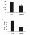
Analysis of cell survival after 2 μM lapatinib treatment. A. A549 cells exposed to 2 μM lapatinib for 24 h exhibited a reduced cell growth proliferation (P < 0.001); B. After 10 days of exposure to lapatinib, the number of colonies was dramatically abrogated (***: P < 0.0001).
Figure 4
Figure 4
Cell cycle study of A549 cells treated with lapatinib. A. Lapatinib significantly alters cell cycle phases (G1 arrest and DNA synthesis reduction) analyzed by flow cytometry after propidium iodide staining (**: P < 0.01; ***: P < 0.001); B. Western blot showing the effect of lapatinib on the expression of the cell cycle regulators cyclins A, B1 and D1. In treated cells, the lower levels of cyclins A and B1, regulators of S and G2/M stages, respectively, corroborated the results described by cytometry. Levels of cyclin D1, a regulator of the G1 phase, were very low and remained unchanged upon treatment.
Figure 5
Figure 5
Intracellular signaling changes induced by lapatinib in A549 cells, analyzed by western blot. A. Immunoblots showing decreased levels of p-EGFR and p-HER-2 after stimulation with 100 ng/ml EGF and treatment with lapatinib. Downstream targets p-AKT, p-ERK1/2, c-Myc and PCNA were also reduced upon exposure to the drug. B. After lapatinib treatment, the proapoptotic protein Bak-1 was increased, the levels of the antiapoptotic proteins IAP-2 and Bcl-xL were reduced, and PARP was cleaved, thus demonstrating that the apoptotic pathway is switched on by this drug in A549 lung cancer cells.
Figure 6
Figure 6
In vivo tumor growth assays. A. After tumor implantation into immunocompromised nude mice, animals were treated with lapatinib for four weeks at the indicated concentration. Tumor volumes in treated mice were smaller than those found in controls; B. Lapatinib significantly reduced tumor metabolism (P = 0.037), which was shown by the standardized glucose uptake values (SUV) measured with micro-PET.
Figure 7
Figure 7
In vivo effect of radiotherapy alone or in combination with lapatinib, in A549 tumor-bearing mice. Lapatinib reduced modestly tumor growth in irradiated animals compared to only irradiated mice.
Figure 8
Figure 8
Lapatinib alters angiogenesis and the number of circulating endothelial progenitors (CEPs) in mice xenotransplanted with A549 tumor cells. A. Representative images of CD31-stained tumors from controls, lapatinib-treated, radiotherapy-treated and radiotherapy plus lapatinib-treated mice. B. Lapatinib dramatically reduced the CD31-positive area in the tumors (*: P < 0.05; **: P < 0.01; ***: P < 0.001); C. Quantification of CEPs in A549 tumor-bearing mice by flow cytometry from the peripheral blood. Lapatinib tended to reduce the number of CEPs compared to controls. Interestingly, it significantly diminished the number of CEPs (P = 0.0167) that were increased after radiotherapy treatment.

Similar articles

Cited by

References

    1. Pajares MJ, Zudaire I, Lozano MD, Agorreta J, Bastarrika G, Torre W, Remirez A, Pio R, Zulueta JJ, Montuenga LM. Molecular profiling of computed tomography screen-detected lung nodules shows multiple malignant features. Cancer Epidemiol Biomarkers Prev. 2006;15:373–380. doi: 10.1158/1055-9965.EPI-05-0320. - DOI - PubMed
    1. Hirsch FR, Scagliotti GV, Langer CJ, Varella-Garcia M, Franklin WA. Epidermal growth factor family of receptors in preneoplasia and lung cancer: perspectives for targeted therapies. Lung Cancer. 2003;41(Suppl 1):S29–S42. doi: 10.1016/S0169-5002(03)00137-5. - DOI - PubMed
    1. Rosell R, Moran T, Queralt C, Porta R, Cardenal F, Camps C, Majem M, Lopez-Vivanco G, Isla D, Provencio M, Insa A, Massuti B, Gonzalez-Larriba JL, Paz-Ares L, Bover I, Garcia-Campelo R, Moreno MA, Catot S, Rolfo C, Reguart N, Palmero R, Sánchez JM, Bastus R, Mayo C, Bertran-Alamillo J, Molina MA, Sanchez JJ, Taron M. Screening for epidermal growth factor receptor mutations in lung cancer. N Engl J Med. 2009;361:958–967. doi: 10.1056/NEJMoa0904554. - DOI - PubMed
    1. Rocha-Lima CM, Soares HP, Raez LE, Singal R. EGFR targeting of solid tumors. Cancer Control. 2007;14:295–304. - PubMed
    1. Nicholson RI, Gee JM, Harper ME. EGFR and cancer prognosis. Eur J Cancer. 2001;37(Suppl 4):S9–15. doi: 10.1016/S0959-8049(01)00231-3. - DOI - PubMed

Publication types

MeSH terms