Expression and activity profiles of DPP IV/CD26 and NEP/CD10 glycoproteins in the human renal cancer are tumor-type dependent
- PMID: 20459800
- PMCID: PMC2876082
- DOI: 10.1186/1471-2407-10-193
Expression and activity profiles of DPP IV/CD26 and NEP/CD10 glycoproteins in the human renal cancer are tumor-type dependent
Abstract
Background: Cell-surface glycoproteins play critical roles in cell-to-cell recognition, signal transduction and regulation, thus being crucial in cell proliferation and cancer etiogenesis and development. DPP IV and NEP are ubiquitous glycopeptidases closely linked to tumor pathogenesis and development, and they are used as markers in some cancers. In the present study, the activity and protein and mRNA expression of these glycoproteins were analysed in a subset of clear-cell (CCRCC) and chromophobe (ChRCC) renal cell carcinomas, and in renal oncocytomas (RO).
Methods: Peptidase activities were measured by conventional enzymatic assays with fluorogen-derived substrates. Gene expression was quantitatively determined by qRT-PCR and membrane-bound protein expression and distribution analysis was performed by specific immunostaining.
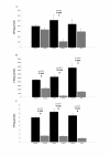
Results: The activity of both glycoproteins was sharply decreased in the three histological types of renal tumors. Protein and mRNA expression was strongly downregulated in tumors from distal nephron (ChRCC and RO). Moreover, soluble DPP IV activity positively correlated with the aggressiveness of CCRCCs (higher activities in high grade tumors).
Conclusions: These results support the pivotal role for DPP IV and NEP in the malignant transformation pathways and point to these peptidases as potential diagnostic markers.
Figures


References
-
- Bannasch P, Zerban H. In: Tumors and tumorlike conditions of the kidneys and ureters. Eble JN, editor. New York: Churchill-Livingstone; 1990. Animal models and renal carcinogenesis; pp. 1–34.
-
- Jin JS, Hsieh DS, Lin YF, Wang JY, Sheu LF, Lee WH. Increasing expression of extracellular matrix metalloprotease inducer in renal cell carcinoma: tissue microarray analysis of immunostaining score with clinicopathological parameters. Int J Urol. 2006;13:573–580. doi: 10.1111/j.1442-2042.2006.01353.x. - DOI - PubMed
-
- Patraki E, Cardillo MR. Quantitative immunohistochemical analysis of matrilysin 1 (MMP-7) in various renal cell carcinoma subtypes. Int J Immunopathol Pharmacol. 2007;20:697–705. - PubMed
Publication types
MeSH terms
Substances
LinkOut - more resources
Full Text Sources
Medical
Research Materials
Miscellaneous

