. 2010;12(3):R81.
doi: 10.1186/ar3006. Epub 2010 May 11.

Altered microRNA expression profile with miR-146a upregulation in CD4+ T cells from patients with rheumatoid arthritis

Affiliations

Altered microRNA expression profile with miR-146a upregulation in CD4+ T cells from patients with rheumatoid arthritis

Jingyi Li et al. Arthritis Res Ther. 2010.

Abstract

Introduction: Increasing evidence indicates that microRNAs (miRNAs) play a critical role in the pathogenesis of inflammatory diseases. The aim of the study was to investigate the expression pattern and function of miRNAs in CD4+ T cells from patients with rheumatoid arthritis (RA).

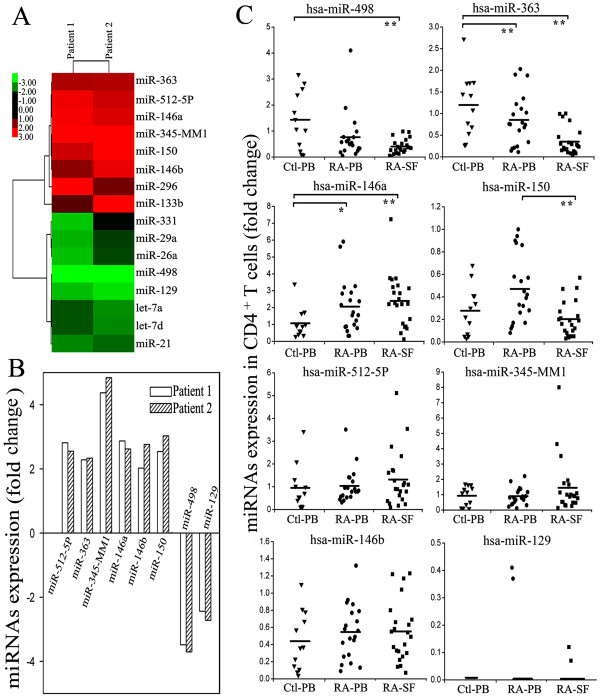
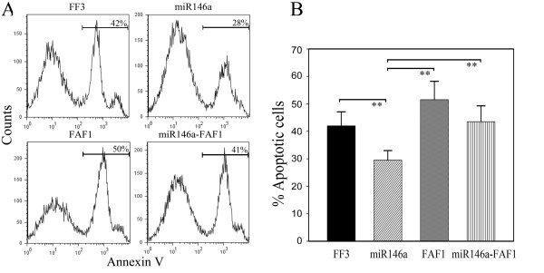
Methods: The expression profile of miRNAs in CD4+ T cells from synovial fluid (SF) and peripheral blood of 33 RA patients was determined by microarray assay and validated by qRT-PCR analysis. The correlation between altered expression of miRNAs and cytokine levels was determined by linear regression analysis. The role of miR-146a overexpression in regulating T cell apoptosis was evaluated by flow cytometry. A genome-wide gene expression analysis was further performed to identify miR-146a-regulated genes in T cells.

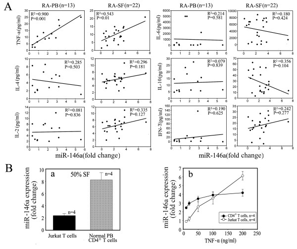
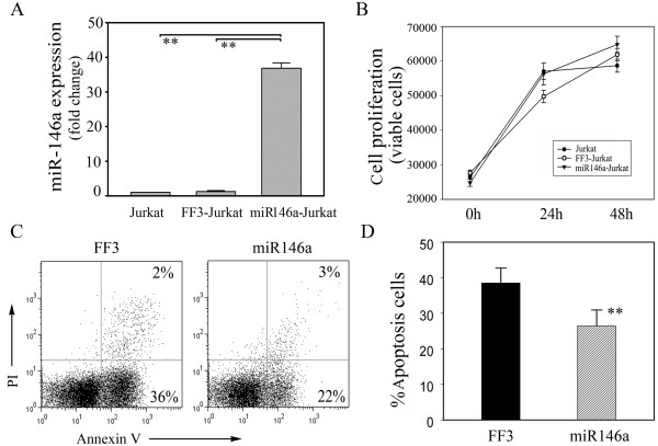
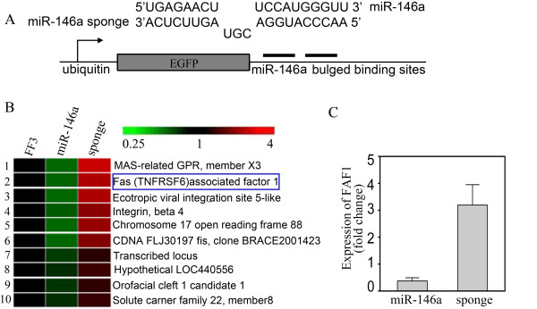
Results: miRNA expression profile analysis revealed that miR-146a expression was significantly upregulated while miR-363 and miR-498 were downregulated in CD4+ T cells of RA patients. The level of miR-146a expression was positively correlated with levels of tumor necrosis factor-alpha (TNF-alpha), and in vitro studies showed TNF-alpha upregulated miR-146a expression in T cells. Moreover, miR-146a overexpression was found to suppress Jurkat T cell apoptosis. Finally, transcriptome analysis of miR-146a overexpression in T cells identified Fas associated factor 1 (FAF1) as a miR-146a-regulated gene, which was critically involved in modulating T cell apoptosis.

Conclusions: We have detected increased miR-146a in CD4+ T cells of RA patients and its close correlation with TNF-alpha levels. Our findings that miR-146a overexpression suppresses T cell apoptosis indicate a role of miR-146a in RA pathogenesis and provide potential novel therapeutic targets.


Figures

Figure 1
Figure 1
The altered expression profile of miRNAs in CD4+ T cells from SF of RA patients. (a) Compared with normal CD4+ T cells, altered expression of 16 microRNAs (miRNAs) with fold change (>2) was found in CD4+ T cells of rheumatoid arthritis (RA) by miRNA microarray analysis. (red: upregulation; green: downregulation) (b) Expression of eight miRNAs was altered in both RA patients by miRNA microarray analysis. Six of them were upregulated and two of them were downregulated. (c) Compared with healthy controls, levels of miRNAs - miR-498, miR-363, miR-146a, miR-150, miR-512-5P, miR-345-MM1, miR-146b and miR-129 - expression in peripheral blood (PB; n = 20) and synovial fluid (SF; n = 22) of RA patients and PB (n = 12) of healthy controls were determined by Taqman quantitative RT-PCR analysis. Horizontal bars represent the mean values. Ctl-PB: CD4+ T cells from PB of healthy controls; RA-PB: CD4+ T cells from PB of RA patients; RA-SF: CD4+ T cells from SF of RA patients. **P < 0.01, *P < 0.05.
Figure 2
Figure 2
Positive correlation of miR-146a expression with TNF-α levels in RA patients. (a) A positive correlation was found between miR-146a expression and TNF-α levels. Of synovial fluid (SF), 50% increased the expression of miR-146a in Jurkat T cells by 2.39 ± 0.34 fold and in human CD4+ T cells by 8.21 ± 1.19 fold (n = 4), respectively. (b) Induction of miR-146a expression stimulated with TNF-α or SF of rheumatoid arthritis (RA) in both Jurkat cells and CD4+ T cells. Unstimulated Jurkat T cells or CD4+ T cells were used as controls, respectively. The value of each control sample was set at one and further used to calculate the fold change. TNF-α upregulated miR-146a expression in Jurkat T cells and human CD4+ T cells in a dose-dependent fashion.
Figure 3
Figure 3
Overexpression of miR-146a suppresses T cell apoptosis. (a) The expression of miR-146a in Jurkat T cells measured by quantitiative RT-PCR increased more than 30 fold after transfection with FUGW-miR-146a, but showed no change after transfection with the control vector FUGW-FF3. (b) After phytohemagglutinin (PHA) stimulation, Jurkat T cells with miR-146a overexpression showed a similar proliferative response as their controls. (c) Upon anti-Fas antibody treatment for six hours, miR-146a-overexpressing Jurkat T cells displayed significantly reduced apoptosis as detected by flow cytometry. (d) After six hours of anti-Fas antibody treatment, apoptotic Jurkat T cells were evaluated. The data are derived from three independent experiments (mean ± standard deviation). **P < 0.01.
Figure 4
Figure 4
Gene expression profiles in Jurkat T cells with up-regulated and down-regulated miR-146a expression. (a) Schematic representation of FUGW-miR-146a sponge, which was inserted into lentivirus vector FUGW. (b) Gene expression profiles of Jurkat T cells transfected with FUGW-miR-146a or FUGW-miR-146a sponge were analyzed by Affymetrix GeneChip. Fas-associated factor 1 (FAF1) was found to be negatively correlated to the level of miR-146a. (c) Expression levels of FAF1 were further verified by quantitative RT-PCR, consistent with the results from gene chip analysis. FUGW-FF3-Jurkat T cells were used as the control. The value of each control sample was set at one and further used to calculate the fold change.
Figure 5
Figure 5
FAF1 abolishes the suppressive effect of miR-146a on Jurkat T cell apoptosis. (a) Fas-induced apoptosis was evaluated in Jurkat T cells transfected with either miR-146a alone or together with Fas-associated factor 1 (FAF1). miR-146a-overexpressing T cells showed significantly reduced apoptosis whereas co-overexpressing FAF1 abrogated the effect of miR-146a on T cell apoptosis. (b) After six hours of anti-Fas treatment, apoptotic Jurkat T cells were evaluated. The data are derived from three independent experiments (mean ± standard deviation). **P < 0.01.

Comment in

References

    1. Panayi GS, Lanchbury JS, Kingsley GH. The importance of the T cell in initiating and maintaining the chronic synovitis of rheumatoid arthritis. Arthritis Rheum. 1992;35:729–735. doi: 10.1002/art.1780350702. - DOI - PubMed
    1. Gregersen PK, Silver J, Winchester RJ. The shared epitope hypothesis. An approach to understanding the molecular genetics of susceptibility to rheumatoid arthritis. Arthritis Rheum. 1987;30:1205–1213. doi: 10.1002/art.1780301102. - DOI - PubMed
    1. Stastny P. Mixed lymphocyte cultures in rheumatoid arthritis. J Clin Invest. 1976;57:1148–1157. doi: 10.1172/JCI108382. - DOI - PMC - PubMed
    1. Ali M, Ponchel F, Wilson KE, Francis MJ, Wu X, Verhoef A, Boylston AW, Veale DJ, Emery P, Markham AF, Lamb JR, Isaacs JD. Rheumatoid arthritis synovial T cells regulate transcription of several genes associated with antigen-induced anergy. J Clin Invest. 2001;107:519–528. doi: 10.1172/JCI8027. - DOI - PMC - PubMed
    1. Silverman HA, Johnson JS, Vaughan JH, McGlamory JC. Altered lymphocyte reactivity in rheumatoid arthritis. Arthritis Rheum. 1976;19:509–515. doi: 10.1002/art.1780190301. - DOI - PubMed

Publication types

MeSH terms