

The promoter for intestinal cell kinase is head-to-head with F-Box 9 and contains functional sites for TCF7L2 and FOXA factors
- PMID: 20459822
- PMCID: PMC2876993
- DOI: 10.1186/1476-4598-9-104
The promoter for intestinal cell kinase is head-to-head with F-Box 9 and contains functional sites for TCF7L2 and FOXA factors
Abstract
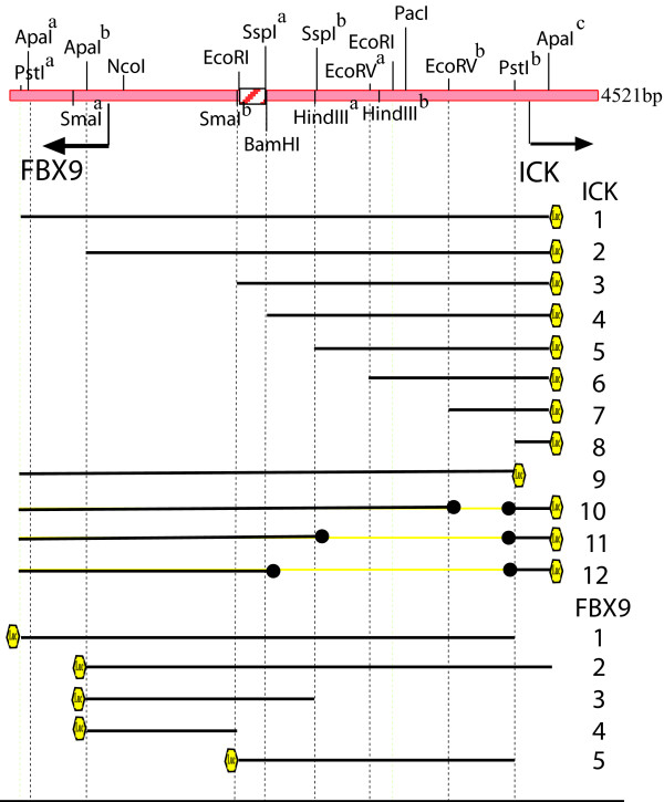
Background: Intestinal cell kinase (ICK; GeneID 22858) is a conserved MAPK and CDK-like kinase that is widely expressed in human tissues. Data from the Cancer Genome Anatomy Project indicated ICK mRNA is increased in cancer, and that its expression correlated with expression of mRNA for an uncharacterized F-box protein, FBX9 (GeneID: 26268). ICK and FBX9 genes are arranged head-to-head on opposite strands, with start sites for transcription separated by approximately 3.3 kb. We hypothesized ICK and FBX9 are potentially important genes in cancer controlled by a bidirectional promoter.
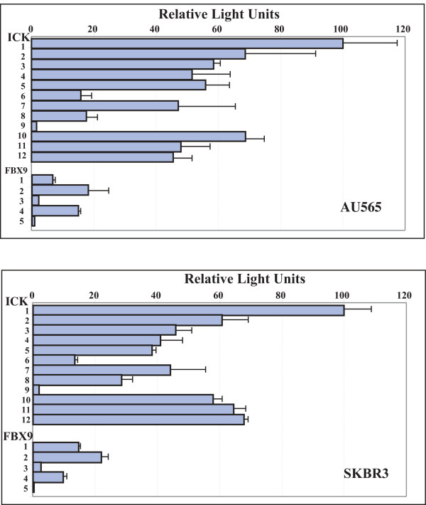
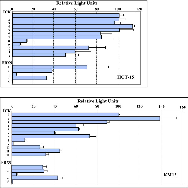
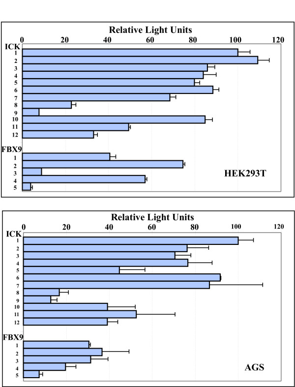
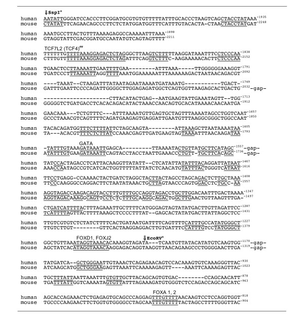
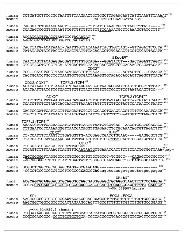
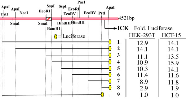
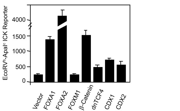
Results: We assessed promoter activity of the intergenic region in both orientations in cancer cell lines derived from breast (AU565, SKBR3), colon (HCT-15, KM12), and stomach (AGS) cancers, as well as in embryonic human kidney (HEK293T) cells. The intergenic segment was active in both orientations in all of these lines, and ICK promoter activity was greater than FBX9 promoter activity. Results from deletions and truncations defined a minimal promoter for ICK, and revealed that repressors and enhancers differentially regulate ICK versus FBX9 promoter activity. The ICK promoter contains consensus motifs for several FOX-family transcription factors that align when mouse and human are compared using EMBOSS. FOXA1 and FOXA2 increase luciferase activity of a minimal promoter 10-20 fold in HEK293T cells. Consensus sites for TCF7L2 (TCF4) (Gene Id: 6934) are also present in both mouse and human. The expression of beta-catenin increased activity of the minimal promoter approximately 10 fold. ICK reference mRNAs (NM_014920.3, NM_016513) are expressed in low copy number and increased in some breast cancers, using a ten base tag 5'-TCAACCTTAT-3' specific for both ICK transcripts.
Conclusion: ICK and FBX9 are divergently transcribed from a bidirectional promoter that is GC-rich and contains a CpG island. A minimal promoter for ICK contains functional sites for beta-catenin/TCF7L2 and FOXA. These data are consistent with functions that have been proposed for ICK in development and in proliferation or survival of some breast and colon cancers.
Figures
References
-
- Fu Z, Schroeder MJ, Shabanowitz J, Kaldis P, Togawa K, Rustgi AK, Hunt DF, Sturgill TW. Activation of a nuclear Cdc2-related kinase within a mitogen-activated protein kinase-like TDY motif by autophosphorylation and cyclin-dependent protein kinase-activating kinase. Mol Cell Biol. 2005;25:6047–6064. doi: 10.1128/MCB.25.14.6047-6064.2005. - DOI - PMC - PubMed
-
- Abe S, Yagi T, Ishiyama S, Hiroe M, Marumo F, Ikawa Y. Molecular cloning of a novel serine/threonine kinase, MRK, possibly involved in cardiac development. Oncogene. 1995;11:2187–2195. - PubMed
-
- Wohlbold L, Larochelle S, Liao JC, Livshits G, Singer J, Shokat KM, Fisher RP. The cyclin-dependent kinase (CDK) family member PNQALRE/CCRK supports cell proliferation but has no intrinsic CDK-activating kinase (CAK) activity. Cell Cycle. 2006;5:546–554. - PubMed
Publication types
MeSH terms
Substances
Grants and funding
LinkOut - more resources
Full Text Sources
Molecular Biology Databases
Miscellaneous
