

Greater lean tissue and skeletal muscle mass are associated with higher bone mineral content in children
- PMID: 20459832
- PMCID: PMC2886077
- DOI: 10.1186/1743-7075-7-41
Greater lean tissue and skeletal muscle mass are associated with higher bone mineral content in children
Abstract
Background: To compare the relationship of skeletal muscle mass with bone mineral content in an ethnically diverse group of 6 to 18 year old boys and girls.
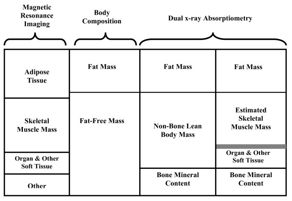
Methods: 175 healthy children (103 boys; 72 girls) had assessments of body mass, height, and Tanner stage. Whole body bone mineral content, non-bone lean body mass (nbLBM), skeletal muscle mass, and fat mass were assessed using dual-energy X-ray absorptiometry (DXA). Muscle mass was estimated from an equation using appendicular lean soft tissue measured by DXA, weight and height. Estimates of skeletal muscle mass and adipose tissue were also assessed by whole body multi-slice magnetic resonance imaging (MRI). Linear regression was used to determine whether skeletal muscle mass assessed by DXA or by MRI were better predictors of bone mineral content compared with nbLBM after adjusting for sex, age, race or ethnicity, and Tanner stage.
Results: Greater skeletal muscle mass was associated with greater bone mineral content (p < 0.001). The skeletal muscle mass assessed by MRI provided a better fitting regression model (determined by R2 statistic) compared with assessment by DXA for predicting bone mineral content. The proportion of skeletal muscle mass in nbLBM was significantly associated with greater bone mineral content adjusted for total nbLBM.
Conclusions: This study is among the first to describe and compare the relationship of skeletal muscle to bone using both MRI and DXA estimates. The results demonstrate that the use of MRI provides a modestly better fitting model for the relationship of skeletal muscle to bone compared with DXA. Skeletal muscle had an impact on bone mineral content independent of total non-bone lean body mass. In addition, Hispanics had greater bone mineral content compared to other race and ethnic groups after adjusting for sex, age, adipose tissue, skeletal muscle mass, and height.
Figures

References
-
- Weinsier RL, Schutz Y, Bracco D. Reexamination of the relationship of resting metabolic rate to fat-free mass and to the metabolically active components of fat-free mass in humans. Am J Clin Nutr. 1992;55:790–794. - PubMed
-
- Ferretti JL, Capozza RF, Cointry GR, Garcia SL, Plotkin H, Alvarez-Figueira ML, Zanchietta JR. Gender-related differences in the relationship between densitometric values of whole body bone mineral content and lean body mass in humans between 2 and 87 years of age. Bone. 1998;22:683–690. doi: 10.1016/S8756-3282(98)00046-5. - DOI - PubMed
LinkOut - more resources
Full Text Sources
Medical
