Comparative Study
. 2010 May 11:10:44.
doi: 10.1186/1471-230X-10-44.

Effective in vivo and ex vivo gene transfer to intestinal mucosa by VSV-G-pseudotyped lentiviral vectors

Affiliations
Comparative Study

Effective in vivo and ex vivo gene transfer to intestinal mucosa by VSV-G-pseudotyped lentiviral vectors

Hiroshi Matsumoto et al. BMC Gastroenterol. .

Abstract

Background: Gene transfer to the gastrointestinal (GI) mucosa is a therapeutic strategy which could prove particularly advantageous for treatment of various hereditary and acquired intestinal disorders, including inflammatory bowel disease (IBD), GI infections, and cancer.

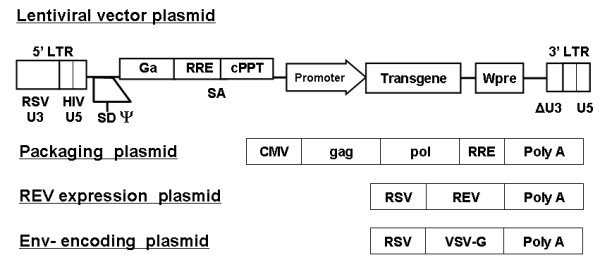
Methods: We evaluated vesicular stomatitis virus glycoprotein envelope (VSV-G)-pseudotyped lentiviral vectors (LV) for efficacy of gene transfer to both murine rectosigmoid colon in vivo and human colon explants ex vivo. LV encoding beta-galactosidase (LV-beta-Gal) or firefly-luciferase (LV-fLuc) reporter genes were administered by intrarectal instillation in mice, or applied topically for ex vivo transduction of human colorectal explant tissues from normal individuals. Macroscopic and histological evaluations were performed to assess any tissue damage or inflammation. Transduction efficiency and systemic biodistribution were evaluated by real-time quantitative PCR. LV-fLuc expression was evaluated by ex vivo bioluminescence imaging. LV-beta-Gal expression and identity of transduced cell types were examined by histochemical and immunofluorescence staining.

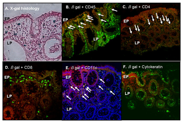
Results: Imaging studies showed positive fLuc signals in murine distal colon; beta-Gal-positive cells were found in both murine and human intestinal tissue. In the murine model, beta-Gal-positive epithelial and lamina propria cells were found to express cytokeratin, CD45, and CD4. LV-transduced beta-Gal-positive cells were also seen in human colorectal explants, consisting mainly of CD45, CD4, and CD11c-positive cells confined to the LP.

Conclusions: We have demonstrated the feasibility of LV-mediated gene transfer into colonic mucosa. We also identified differential patterns of mucosal gene transfer dependent on whether murine or human tissue was used. Within the limitations of the study, the LV did not appear to induce mucosal damage and were not distributed beyond the distal colon.


Figures

Figure 1
Figure 1
Lentiviral vector (LV) constructs and ex vivo explant culture system. (A) Schematic representation of self-inactivating vectors containing a central polypurine tract (cPPT)/central termination sequence, immediate early cytomegalovirus (CMV) promoter. Vectors were constructed for expression of beta-galactosidase (β-Gal) and firefly luciferase (fLuc).
Figure 2
Figure 2
In vitro transduction of vesicular stomatitis virus G protein (VSV-G)-pseudotyped lentivirus (LV) encoding β-Gal in colonic cell lines. (A) Percentage of cells transduced by the VSV-G-pseudotyped LV. One hundred cells were counted in three randomly selected, non-adjacent fields in triplicate. (B) Colon adenocarcinoma cell lines showing evidence of β-Gal transduction following exposure to 10 ng p24 of vector. All values were expressed as means ± SD. *p < 0.05 compared with the results of 0.1 ng p24 VSV-G LV transduction.
Figure 3
Figure 3
Bioluminescence imaging analysis. (A) Ex vivo bioluminescence imaging analysis after rectal administration of vesicular stomatitis virus-G protein (VSV-G)-pseudotyped lentivirus (LV) expressing firefly-luciferase (fLuc). This pseudocolor image, which is superimposed on a grayscale reference image, uses color (blue, least intense; red, most intense) to illustrate signal strength. (B) Photon emission (p/s/cm2/sr) in the region of interest (ROI) of colon transduced by VSVG-pseudotyped LV shows significantly higher levels of transduction compared to the mock control. (C) Biodistribution of VSV-G LV after rectal administration demonstrated by real-time quantitative polymerase chain reaction (qPCR) analysis of viral copy number in 300 ng of genomic DNA of in vivo (equivalent to approximately 50,000 cells). The detection limit was 50 copies per 300 ng genomic DNA.
Figure 4
Figure 4
X-Gal histology in murine colon transduced by vesicular stomatitis virus G protein (VSV-G)-pseudotyped lentivirus (LV) encoding β-Gal in vivo. The time course of β-Gal expression in the colon of BALB/c mice. Each group had 6 mice. The colon was removed at the indicated time points. The number of X-Gal-positive cells in murine colon transduced by LV-β-Gal in vivo. All values were expressed as means ± SD. *p < 0.05 compared with the results of normal control (NC).
Figure 5
Figure 5
Double immunofluorescence histology analysis in murine colon. (A) Histochemical analysis of transduced murine colon in vivo. The X-Gal-positive cells were evident mainly on the luminal surface of the distal colon (magnification × 400). (B-F) Double immunofluorescence staining of tranduced murine colon examined under confocal microscopy. On Day 2 following rectal administration, β-Gal-positive cells in murine colon tissue expressed CD45, CD4, and cytokeratin, but not CD8 or CD11c. In these photomicrographs, β-Gal-positive cells are visualized by red fluorescence, CD phenotypes and cytokeratin are visualized by green fluorescence, and all images display fluorescence overlays: (B) β-Gal and CD45; (C) β-Gal and CD4; (D) β-Gal and CD8; (E) β-Gal and CD11c; and, (F) β-Gal and cytokeratin. Double-positive cells indicated by the arrowheads are yellow. (EP; epithelial cell, LP; lamina propria cell)
Figure 6
Figure 6
X-Gal histology and double immunofluorescence histology analysis in human colonic explants transduced by VSV-G-pseudotyped lentivirus (LV) encoding β-Gal. (A) Histological appearance of transduced human explants after X-Gal staining. X-Gal-positive cells, here stained blue, were found mainly in the LP. (B-F) Double immunofluorescence confocal microscopy of human explants transduced with VSV-G. β-Gal-positive cells expressed CD4, CD45, and CD11c, but not CD8, or cytokeratin. In these photomicrographs, β-Gal is visualized by red fluorescence, each cellular phenotype marker is visualized by green fluorescence, and nuclei are visualized by blue DAPI staining, respectively, and all images display fluorescence overlays: (B) β-Gal and CD45; (C) β-Gal and CD4; (D) β-Gal and CD8; (E) β-Gal and CD11c; and, (F) β-Gal and cytokeratin. Double-positive cells, indicated by the arrowheads, are yellow (EP; epithelial cell, LP; lamina propria cell).

Similar articles

Cited by

References

    1. Prieto J, Herraiz M, Sangro B, Qian C, Mazzolini G, Melero I. et al.The promise of gene therapy in gastrointestinal and liver diseases. Gut. 2003;52(Suppl 2):ii49–ii54. - PMC - PubMed
    1. Raper SE, Wilson JM. Making space for intestinal gene therapy. Gastroenterology. 1997;112:1753–1756. doi: 10.1016/S0016-5085(97)70060-X. - DOI - PubMed
    1. Forbes SJ, Hodgson HJ. Review article: gene therapy in gastroenterology and hepatology. Aliment Pharmacol Ther. 1997;11:823–836. doi: 10.1046/j.1365-2036.1997.00226.x. - DOI - PubMed
    1. van MC, Te Velde AA, venter SJ van De, Rodriguez Pena MS. Gene therapy in the treatment of intestinal inflammation. Int J Colorectal Dis. 2004;19:79–86. doi: 10.1007/s00384-003-0501-4. - DOI - PubMed
    1. Wirtz S, Neurath MF. Gene transfer approaches for the treatment of inflammatory bowel disease. Gene Ther. 2003;10:854–860. doi: 10.1038/sj.gt.3302013. - DOI - PubMed

Publication types