. 2010 Sep;157(1):55-62.
doi: 10.1016/j.cbpa.2010.05.002. Epub 2010 May 10.

Physiological and pharmacological characterizations of the larval Anopheles albimanus rectum support a change in protein distribution and/or function in varying salinities

Affiliations

Physiological and pharmacological characterizations of the larval Anopheles albimanus rectum support a change in protein distribution and/or function in varying salinities

Kristin E Smith et al. Comp Biochem Physiol A Mol Integr Physiol. 2010 Sep.

Abstract

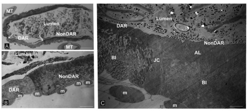
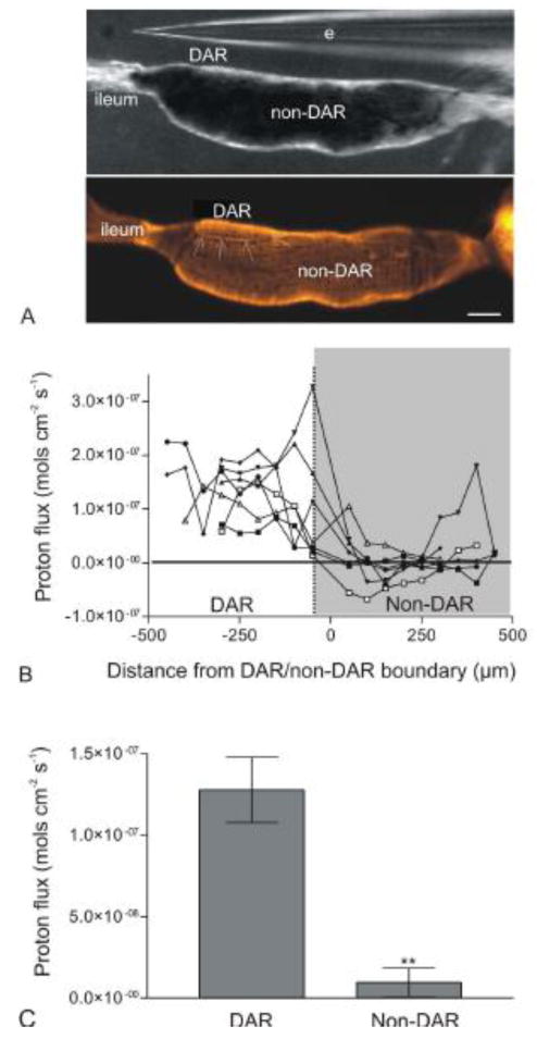
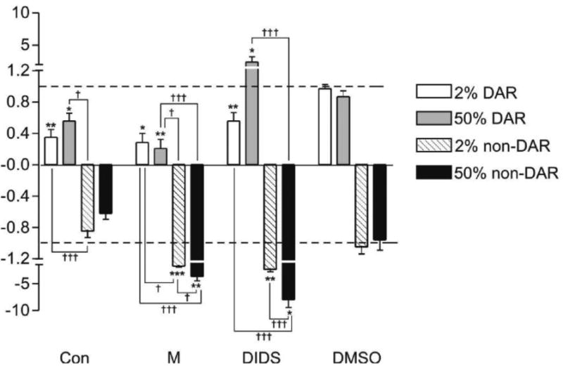
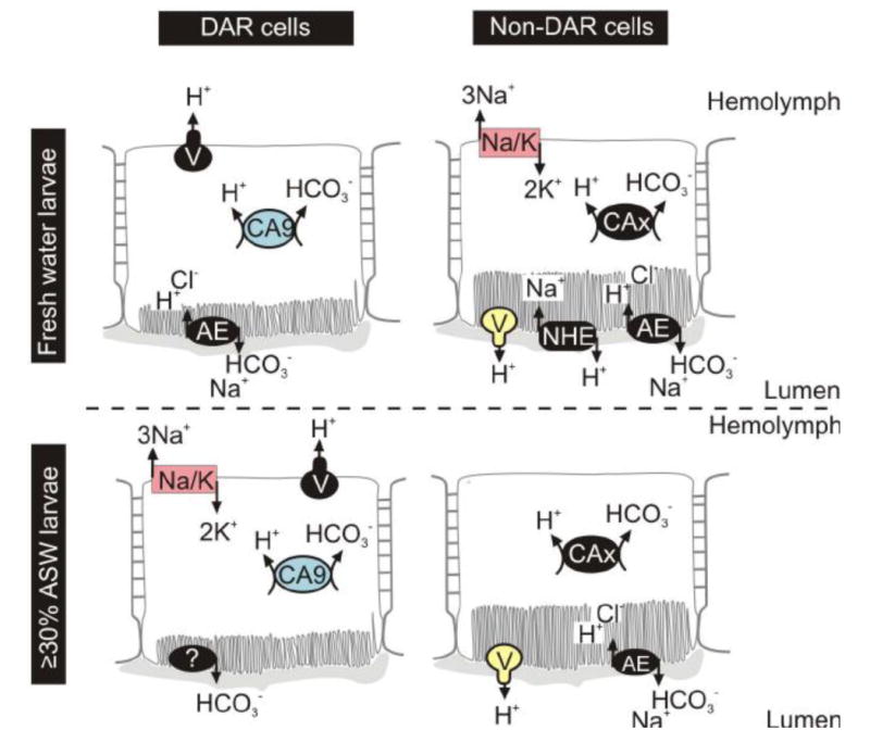
Ion regulation is a biological process crucial to the survival of mosquito larvae and a major organ responsible for this regulation is the rectum. The recta of anopheline larvae are distinct from other subfamilies of mosquitoes in several ways, yet have not yet been characterized extensively. Here we characterize the two major cell types of the anopheline rectum, DAR and non-DAR cells, using histological, physiological, and pharmacological analyses. Proton flux was measured at the basal membrane of 2%- and 50%-artificial sea water-reared An. albimanus larvae using self-referencing ion-selective microelectrodes, and the two cell types were found to differ in basal membrane proton flux. Additionally, differences in the response of that flux to pharmacological inhibitors in larvae reared in 2% versus 50% ASW indicate changes in protein function between the two rearing conditions. Finally, histological analyses suggest that the non-DAR cells are structurally suited for mediating ion transport. These data support a model of rectal ion regulation in which the non-DAR cells have a resorptive function in freshwater-reared larvae and a secretive function in saline water-reared larvae. In this way, anopheline larvae may adapt to varying salinities.


Figures

Figure 1
Figure 1
Rectal cytology. The rectal cells of freshwater-reared An. gambiae larvae are lined on the luminal (apical) side by cuticle and on the hemolymph barrier (basal) side by circumferential muscles. Two cell types are distinguishable by the presence or absence of a prominent apical border of apical lamellae (AL, panels “B” and “C”). Panel “A” shows relatively low magnification and the disposition of the DAR and nonDAR cells relative to the borders between the two cell types (arrows) in the anterior region of the rectum and the surrounding profiles of Malpighian tubules (MT). Panel “B” is a higher magnification of the boundary of the two cell types with the basal array of muscle fiber bundles (m) indicated. Note the clear distinction in appearance of the DAR and non-DAR cells at the luminal (apical) surface. Panel “C” shows a transmission electron micrograph of the DAR-nonDAR junction depicting the disparity in the organization of the two cell types. Also evident is a third cell type which is a junctional cell (JC) wich separates the DAR and non DAR cells. Also note that the appearance of the elaboration of basal membranes known as the basal infoldings (BI) is distinct between the DAR and nonDAR cells. Circumferential muscle fiber bundles at the basal extreme (m) are also evident. Magnification bars: “A” = 50 μm; “B” = 12.5 μm; “C” = 3.6 μm.
Figure 2
Figure 2
Measure of proton flux at the basal membrane of the DAR and non-DAR cells of An. albimanus larvae using ISMs in a self-referencing mode. (A) Image illustrating the larval rectum under a light microscope during proton flux measurements (top panel), and post-stained with rhodamine 123 to visualize the DAR/non-DAR boundary (arrows; bottom panel). (B) Proton flux measurements for seven individual preparations (three larvae reared in 2% ASW and four larvae reared in 50% ASW) beginning at the rectum-ileum junction and continuing in 50μm intervals until the end of the rectal cells. Each data point represents an average of continuous measurements taken over a five minute period. There was a consistent, large proton efflux in the area of the DAR cells in all preparations. This efflux dropped drastically, often becoming an influx at the non-DAR cells. (C) Averaged DAR and non-DAR proton flux measurements of the same seven larvae represented in (B). Error bars indicate standard error of the mean. Difference in proton flux between the DAR and non-DAR cells was statistically significant (P-value < 0.01) as determined using a paired t-test and reported as a two-tailed P-value. Scale bar: 100μm. e: electrode. □, ●, ○: larvae reared in 2% ASW; ◆, ▼, ▲, ■: larvae reared in 50% ASW.
Figure 3
Figure 3
Pharmacological inhibition of proton flux at the basal membrane of 2% and 50% ASW-reared An. albimanus rectum. Y-axis indicates ratio of flux after treatment, to flux before treatment with an inhibitor. A value of “1” (for efflux) or “-1” (for influx) indicates no change in flux and is indicated by a dashed line. Inhibitors included a V-ATPase inhibitor, concanamycin A (Con; 10-6 mol L-1), a carbonic anhydrase inhibitor, methazolamide (M; 10-4 mol L-1), and an anion exchange inhibitor, DIDS (10-6 mol L-1), as well as a DMSO (10-6 mol L-1) vehicle control. All results represent an average of n > 5 preparations. Significance asterisks: * = P-value < 0.05; ** = P-value < 0.01; *** = P-value < 0.005. Asterisks represent significance of flux before, to flux after inhibitor for each individual preparation. Significance daggers: † = P-value < 0.05; ††= P-value < 0.01; ††† = P-value < 0.001. Daggers represent significance of flux inhibition between cell types and rearing conditions for each inhibitor.
Figure 4
Figure 4
Putative model for ion transport in the anopheline rectum of larvae reared in fresh or saline water. Proteins in color have been previously immunolocalized, whereas proteins in black are predicted. AE: anion exchanger; CA: carbonic anhydrase; Na/K: sodium/potassium ATPase; NHE: sodium/hydrogen exchanger; V: V-ATPase.

Similar articles

Cited by

References

    1. Beyenbach KW. Mechanism and regulation of electrolyte transport in Malpighian tubules. J Insect Physiol. 1995;4:197–207.
    1. Boron WF. Sodium-coupled bicarbonate transporters. JOP. 2001;2:176–181. - PubMed
    1. Bradley TJ. Evidence for hypo- and hyperosmotic regulation in the larvae of an anopheline mosquito. Am Zool. 1987a;27(4):30A.
    1. Bradley TJ. Physiology of osmoregulation in mosquitoes. Annu Rev Entomol. 1987b;32:439–462. - PubMed
    1. Bradley TJ. The role of physiological capacity, morphology, and phylogeny in determining habitat use in mosquitoes. In: Wainwright PC, Reilly SM, editors. Ecological Morphology. The University of Chicago Press; Chicago and London: 1994. pp. 303–318.

Publication types

MeSH terms

LinkOut - more resources