Review
. 2010:281:91-128.
doi: 10.1016/S1937-6448(10)81003-2.

New insights into the regulation of the actin cytoskeleton by tropomyosin

Affiliations
Review

New insights into the regulation of the actin cytoskeleton by tropomyosin

C-L Albert Wang et al. Int Rev Cell Mol Biol. 2010.

Abstract

The actin cytoskeleton is regulated by a variety of actin-binding proteins including those constituting the tropomyosin family. Tropomyosins are coiled-coil dimers that bind along the length of actin filaments. In muscles, tropomyosin regulates the interaction of actin-containing thin filaments with myosin-containing thick filaments to allow contraction. In nonmuscle cells where multiple tropomyosin isoforms are expressed, tropomyosins participate in a number of cellular events involving the cytoskeleton. This chapter reviews the current state of the literature regarding tropomyosin structure and function and discusses the evidence that tropomyosins play a role in regulating actin assembly.


Figures

Figure 3.1
Figure 3.1
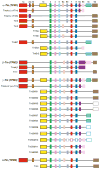
Schematic of exon organization of the four tropomyosin genes in mammals where exons are represented by boxes and introns are represented by solid lines. Isoform variation is primarily at the amino and carboxyl ends and a result, for example, of the alternative use of exon 2a versus 2b in TPM1 and one of the four exons (9a, 9b, 9c, and 9d) at the C-terminus. The figure is based on previously published figures (Gunning et al., 2008; Lin et al., 2008; Vrhovski et al., 2008).
Figure 3.2
Figure 3.2
Domain structures of smooth muscle-specific CaD (h-CaD) and nonmuscle CaD (l-CaD). Both isoforms are derived from the same gene via alternative splicing. In l-CaD a highly charged repeating sequence, which forms a helical stretch in the middle of the h-CaD molecule, is missing, while the other functional domains are identical for the two isoforms.
Figure 3.3
Figure 3.3
Major phosphorylation sites in the C-terminal actin-binding region of CaD. The sequence near the C-terminal end of CaD encompasses the sites for ERK (497, 527)- and PAK (452, 482)- mediated phosphorylation. The numbers for the residues in the human l-CaD sequence are shown here. The numbers in parentheses relate to the corresponding residues in the h-CaD sequence. Note that the two phosphorylation sites in each set are separated by the same number (30) of residues.
Figure 3.4
Figure 3.4
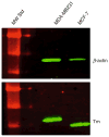
Metastatic and nonmetastatic human breast tumor cells contain different Tms. Western blot analysis with Odyssey software showing that the Tm in the two types of tumor cells, MDA-MB231 (metastatic) and MCF-7 (nonmetastatic), exhibit different mobilities on SDS–polyacrylamide gels (lower panel). β-Actin was used as a reference for loading (upper panel).

References

    1. Alahyan M, Webb MR, Marston SB, El-Mezgueldi M. The mechanism of smooth muscle caldesmon–tropomyosin inhibition of the elementary steps of the acto-myosin ATPase. J Biol Chem. 2006;281:19433–19448. - PubMed
    1. Ansari S, Alahyan M, Marston SB, El-Mezgueldi M. Role of caldesmon in the Ca2+ regulation of smooth muscle thin filaments: evidence for a cooperative switching mechanism. J Biol Chem. 2008;283:47–56. - PubMed
    1. Bailey K. Tropomyosin: a new asymmetric protein component of the muscle fibril. Biochem J. 1948;43:271–279. - PMC - PubMed
    1. Beisel KW, Kennedy JE. Identification of novel alternatively spliced isoforms of the tropomyosin-encoding gene, TMnm, in the rat cochlea. Gene. 1994;143:251–256. - PubMed
    1. Berg JS, Powell BC, Cheney RE. A millennial myosin census. Mol Biol Cell. 2001;12:780–794. - PMC - PubMed

Publication types

MeSH terms

LinkOut - more resources