. 2010 Jun;61(11):2951-65.
doi: 10.1093/jxb/erq119. Epub 2010 May 11.

Petunia nectar proteins have ribonuclease activity

Affiliations

Petunia nectar proteins have ribonuclease activity

Melissa S Hillwig et al. J Exp Bot. 2010 Jun.

Abstract

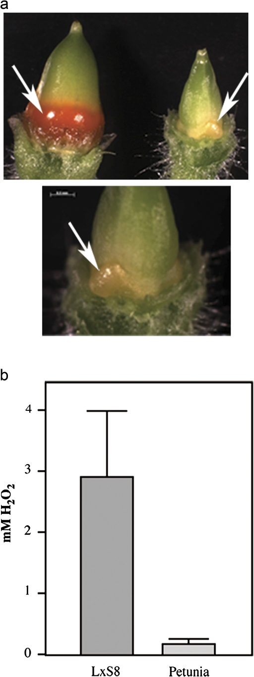
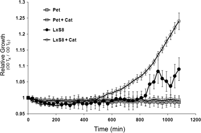
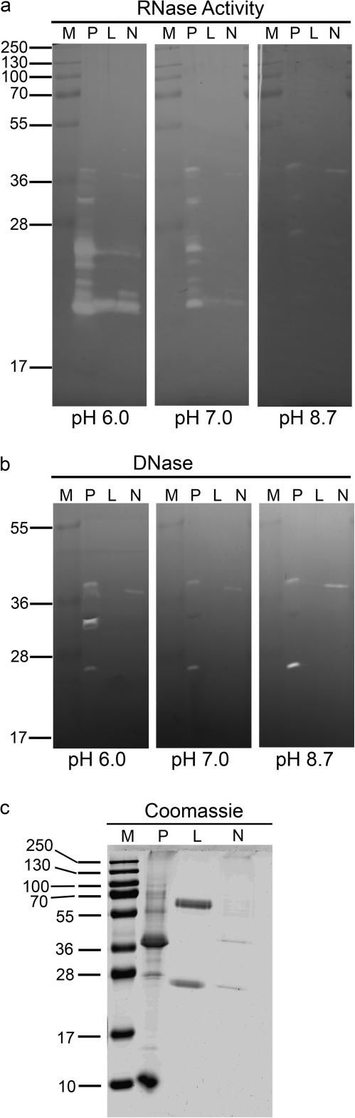
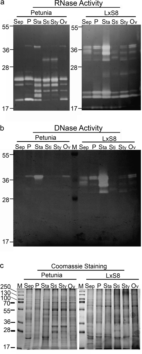
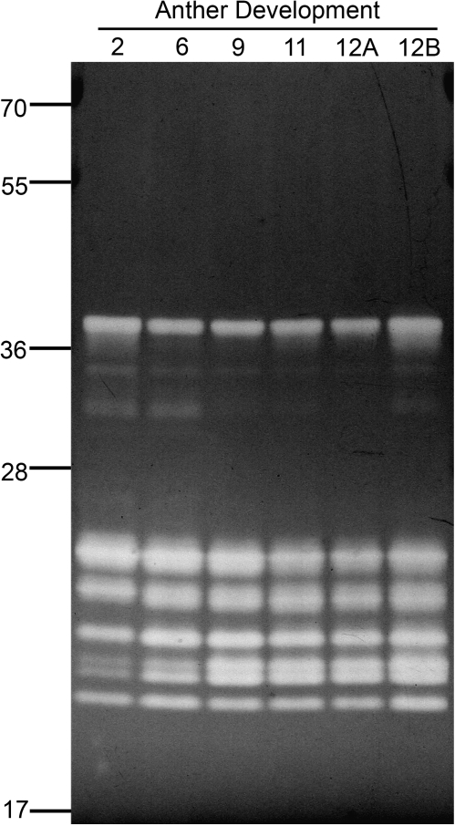
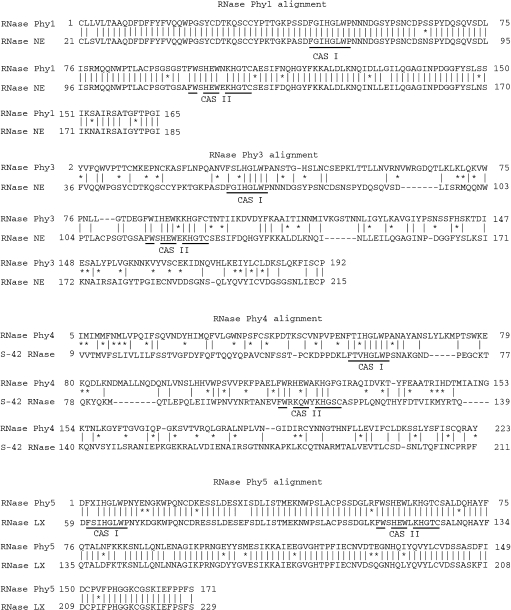
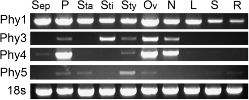
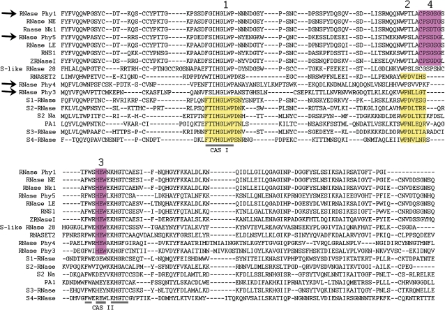
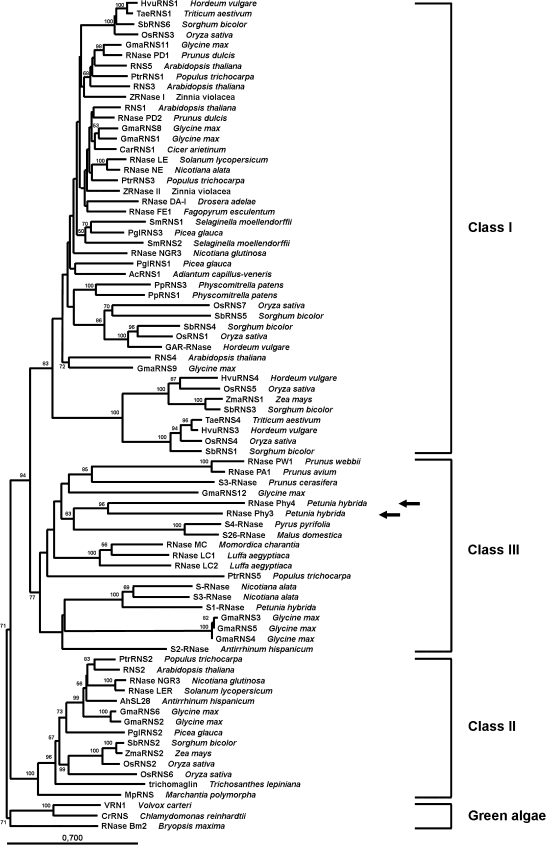
Plants requiring an insect pollinator often produce nectar as a reward for the pollinator's visitations. This rich secretion needs mechanisms to inhibit microbial growth. In Nicotiana spp. nectar, anti-microbial activity is due to the production of hydrogen peroxide. In a close relative, Petunia hybrida, limited production of hydrogen peroxide was found; yet petunia nectar still has anti-bacterial properties, suggesting that a different mechanism may exist for this inhibition. The nectar proteins of petunia plants were compared with those of ornamental tobacco and significant differences were found in protein profiles and function between these two closely related species. Among those proteins, RNase activities unique to petunia nectar were identified. The genes corresponding to four RNase T2 proteins from Petunia hybrida that show unique expression patterns in different plant tissues were cloned. Two of these enzymes, RNase Phy3 and RNase Phy4 are unique among the T2 family and contain characteristics similar to both S- and S-like RNases. Analysis of amino acid patterns suggest that these proteins are an intermediate between S- and S-like RNases, and support the hypothesis that S-RNases evolved from defence RNases expressed in floral parts. This is the first report of RNase activities in nectar.


Figures

Fig. 1.
Fig. 1.
Differences in nectary appearance and nectar composition between petunia and tobacco. (a) Appearance of petunia (right in upper panel, and lower panel) and the L×S8 tobacco hybrid (left, upper panel) nectaries (arrows) from flowers at stage 12 (Koltunow et al., 1990). Observe the differences in size and colour; small, light yellow nectaries in petunia, large, bright orange nectaries in tobacco. (b) Accumulation of hydrogen peroxide in petunia and tobacco nectar. Nectar collected from at least 20 different flowers was pooled and analysed for the presence of H2O2 using a colorimetric assay.
Fig. 2.
Fig. 2.
Effect of tobacco (circles) and petunia (boxes) nectar on the growth of bacteria. Growth of Pseudomonas fluorescens (strain A506) in raw nectar (filled symbols) or nectar that was preincubated with catalase (empty symbols) was followed by changes in OD. Each point represents the mean ±SD (n=3). Data are representative of two independent experiments.
Fig. 3.
Fig. 3.
Nuclease activities are present in nectar. (a) Aliquots (50 μl) of raw nectar from Petunia hybrida and two different tobaccos (Nicotiana tabacum cv. Xanthi and the hybrid Nicotiana langsdorffii×Nicotiana sanderae var. L×S8) were analysed in an in gel RNase activity assay at three different pHs. P, petunia; L, L×S8; N, Xanthi. Size (kDa) of molecular weight markers (M) is indicated. (b) Same samples as in (a), but analysed in an in gel DNase activity assay. (c) The same samples as in (a), analysed by SDS-PAGE, and stained with Coomassie Blue. Gels are representative of at least three independent experiments.
Fig. 4.
Fig. 4.
Nuclease profiles of different floral parts of petunia and ornamental tobacco plants. Flowers were harvested at stage 12, and dissected to obtain sepals (Sep), petals (P), stamens (Sta), stigmas (Sti), styles (Sty), and ovaries (including nectaries, Ov). Total protein extracts (100 μg) from each floral part were analysed in an in gel RNase activity assay (a) or DNase activity assay (b) at pH 6.0. (c) Same samples as in (a) analysed by SDS-PAGE, and stained with Coomassie Blue. Position of molecular weight markers (kDa) is indicated. Gels are representative of at least three independent experiments.
Fig. 5.
Fig. 5.
RNase profile of petunia stamens during development. Stamens were collected from flowers at pre-dehiscence (2, 6, 9, 11, 12A) and post-dehiscence (12B) stages. Total protein extracts (100 μg) were analysed in an in gel RNase activity assay at pH 7.0. Position of molecular weight markers (kDa) is indicated. Gel is representative of at least three independent experiments.
Fig. 6.
Fig. 6.
Petunia RNases have homology to RNase T2 enzymes from other plants. BLAST analysis of predicted RNases encoded by petunia cDNAs amplified from ovaries and nectaries RNA. Alignment of each petunia RNase (RNase Phy1, RNase Phy3, RNase Phy4, and RNase Phy5) with the homologue with the highest BLAST score is shown.
Fig. 7.
Fig. 7.
Expression of petunia RNases in different flower parts. Flowers were harvested at stage 12, and dissected to obtain sepals (Sep), petals (P), stamens (Sta), stigmas (Sti), styles (Sty), ovaries (including nectaries, Ov), and nectaries (N). At the same time, leaves (L), stems (S), and roots (R) were collected. Expression of the four RNase genes was analysed by RT-PCR. Amplification of 18S was used as control for loading. Gels are representative of at least three independent experiments.
Fig. 8.
Fig. 8.
Presence of S- and S-like RNase-specific patterns (according to (Vieira et al., 2008)) in petunia RNases. Alignment of the petunia RNases and representative members of the S-RNase and the S-like RNase subfamilies. Patterns 1 and 2 that define S-RNases are highlighted in yellow; S-like RNase patterns are pink. The conserved active sites (CAS) I and II, typical of RNase T2 enzymes, are indicated. Petunia RNases are indicated with arrows. Accession number of other S-like RNase proteins in the alignment are AAA21135 (RNase NE), BAA95448 (RNase Nk1), X79337 (RNase LE), P42813 (RNS1), AAC49325 (ZRNaseII), CAC50874 (S-like RNase 28); S-RNases included are BAA83479 (S1-RNase), CAA65319 (S2-RNase), AAB40027 (S2 Na), BAD11006 (PA1), AAB07492 (S3-RNase), and BAA28354 (S4-RNase). We also included NP_003721 (RNASET2) from Homo sapiens.
Fig. 9.
Fig. 9.
Phylogenetic relationship of plant RNase T2 proteins. The Neighbor–Joining tree was estimated using only conserved regions of plant RNase T2 proteins. Bootstraps percentages greater than 50 are shown on interior branches. The tree was rooted using algae sequences. Classes I, II, and II clades are indicated, as well as algae proteins. Accession numbers of proteins included in the tree are those described in MacIntosh et al. (2010), with the addition of RNase Phy3 (arrow), RNase Phy4 (arrow), RNase PW1 (ABY86422), RNase PA1 (BAD11006), S3-RNase from P. cerasifera (CAN90133), S4-RNase (BAA28354), S26-RNase (AAB70515), S-RNase (BAA24017), S3-RNase from N. alata (AAB07492), S1-RNase (AAA60465), and S2-RNase (CAA65319).

References

    1. Ai Y, Tsai D-S, Kao T-h. Cloning and sequencing of cDNAs encoding two SSS. Plant Molecular Biology. 1992;19:523–528. - PubMed
    1. Banovic B, Surbanovski N, Konstantinovic M, Maksimovic V. Basic RNases of wild almond (Prunus webbii): cloning and characterization of six new S-RNase and one ‘non-S RNase’ genes. Journal of Plant Physiology. 2009;166:395–402. - PubMed
    1. Bariola P, Green P. Plant ribonucleases. In: D'Alessio G, Riordan J, editors. Ribonucleases: structures and functions. New York: Academic Press; 1997. pp. 163–190.
    1. Bariola PA, Howard CJ, Taylor CB, Verburg MT, Jaglan VD, Green PJ. The Arabidopsis ribonuclease gene RNS1 is tightly controlled in response to phosphate limitation. The Plant Journal. 1994;6:673–685. - PubMed
    1. Bariola PA, MacIntosh GC, Green PJ. Regulation of S-like ribonuclease levels in arabidopsis. Antisense inhibition of RNS1 or RNS2 elevates anthocyanin accumulation. Plant Physiology. 1999;119:331–342. - PMC - PubMed

Publication types