. 2010 Jul 16;285(29):22299-307.
doi: 10.1074/jbc.M109.098566. Epub 2010 May 11.

Loss of cystic fibrosis transmembrane conductance regulator function enhances activation of p38 and ERK MAPKs, increasing interleukin-6 synthesis in airway epithelial cells exposed to Pseudomonas aeruginosa

Affiliations

Loss of cystic fibrosis transmembrane conductance regulator function enhances activation of p38 and ERK MAPKs, increasing interleukin-6 synthesis in airway epithelial cells exposed to Pseudomonas aeruginosa

Julie Bérubé et al. J Biol Chem. .

Abstract

In cystic fibrosis (CF), the absence of functional cystic fibrosis transmembrane conductance regulator (CFTR) translates into chronic bacterial infection, excessive inflammation, tissue damage, impaired lung function and eventual death. Understanding the mechanisms underlying this vicious circle of inflammation is important to design better therapies for CF. We found in CF lung biopsies increased immunoreactivity for p38 MAPK activity markers. Moreover, when compared with their non-CF counterpart, airway epithelial cells expressing the most common mutation in CF (CFTRDeltaF508) were more potent at inducing neutrophil chemotaxis through increased interleukin (IL)-6 synthesis when challenged with Pseudomonas aeruginosa diffusible material. We then discovered that in CFTRDeltaF508 cells, the p38 and ERK MAPKs are hyperactivated in response to P. aeruginosa diffusible material, leading to increased IL-6 mRNA expression and stability. Moreover, although TLR5 contributes to p38 MAPK activation upon P. aeruginosa challenge, it only played a weak role in IL-6 synthesis. Instead, we found that the production of reactive oxygen species is essential for IL-6 synthesis in response to P. aeruginosa diffusible material. Finally, we uncovered that in CFTRDeltaF508 cells, the extracellular glutathione levels are decreased, leading to a greater sensitivity to reactive oxygen species, providing an explanation for the hyperactivation of the p38 and ERK MAPKs and increased IL-6 synthesis. Taken together, our study has characterized a mechanism whereby the CFTRDeltaF508 mutation in airway epithelial cells contributes to increase inflammation of the airways.


Figures

FIGURE 1.
FIGURE 1.
p38 MAPK activation in the airway epithelium of CF patients. Phosphorylated HSP27 (phospho-Ser82) and MAPKAP-K2 (phospho-Thr334) were detected in tissue sections from a healthy adult (Non CF) and a CF patient lung (CF) by immunohistochemistry. Representative fields at higher magnification are shown as insets. Isotype control is presented in the right panels. Pictures are representative of two independent experiments.
FIGURE 2.
FIGURE 2.
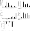
p38 MAPK-dependent IL-6 synthesis is greatly enhanced in AECs expressing CFTRΔF508 in response to P. aeruginosa filtrates. NuLi (non-CF) and CuFi (CFTRΔF508) AECs were left untreated (white bars) or exposed to P. aeruginosa (PA) filtrates in the absence (light gray bars) or presence of BIRB0796 (0.1 μm; dark gray bars) or SB203580 (5 μm; black bars) for 24 h. Medium was collected and incubated with a RayBio Human Inflammation Antibody Array 3 to screen for 40 different inflammatory proteins (A). The amount of fluorescence for each spot was quantified using a Licor Odyssey imaging system. The p38 MAPK-dependent genes are shown in B. GM-CSF, granulocyte-macrophage colony-stimulating factor; RANTES, regulated on activation normal T cell expressed and secreted. Error bars, S.D.
FIGURE 3.
FIGURE 3.
IL-6 secretion by AECs expressing CFTRΔF508 stimulate neutrophil migration. A, neutrophil chemotaxis was assayed in the presence of AEC-conditioned medium without stimulation (Ctl) or exposed to P. aeruginosa filtrates for 3 h (PAF). The contribution of IL-6 was determined by the addition of a neutralizing antibody (aIL-6; 50 μg/ml) to the conditioned medium. B, to test the direct effect of IL-6 on neutrophil migration, recombinant human IL-6 was added in the lower chamber instead of the AECs conditioned medium and the experiment was performed as in A. *, p < 0.05, compared with control (Ctl); #, p < 0.05, compared with P. aeruginosa stimulation. Error bars, S.D.
FIGURE 4.
FIGURE 4.
IL-6 mRNA abundance and stability is increased in P. aeruginosa (PA) filtrates stimulated-CFTRΔF508 AECs. NuLi (non-CF; white bars) and CuFi (CFTRΔF508; black bars) AECs were left untreated or pretreated for 1 h with 0.1 μm BIRB0796 (B, D, and E) or 2 μm PD184352 (B and D) and exposed to P. aeruginosa filtrates for increasing periods of time (A and C), 1 h (B and E), 3 h (D), or 24 h (E). The amount of IL-6 mRNA (A, B, and E) was determined by semiquantitative real-time PCR and the presence of IL-6 in the supernatant (C and D) quantified by ELISA. E, alternatively, following stimulation, 5 μg/ml actinomycin D was added to block transcription, and the RNA was extracted at various intervals thereafter from 0.5 to 6 h. The rates of degradation were calculated over the time course. A rate of 1 would be a completely stable messenger RNA. Representative results of three independent experiments are shown. ¶, p < 0.05; ¶¶, p < 0.01; ¶¶¶, p < 0.001, Cufi compared with Nuli; ##, p < 0.01; ###, p < 0.001, compared with P. aeruginosa stimulation. Error bars, S.D.
FIGURE 5.
FIGURE 5.
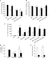
P. aeruginosa filtrate stimulation of CFTRΔF508 AECs leads to enhanced activation of p38 and ERK MAPK pathways. NuLi (non-CF, gray lines) and CuFi (CFTRΔF508, black lines) AECs were left untreated or exposed to P. aeruginosa (PA) filtrates for increasing periods of time as indicated. Following stimulation, cells were lysed, and 20 μg of Triton-soluble materials was subjected to SDS-PAGE. After transfer to nitrocellulose, the membranes were probed with antibodies recognizing only the phosphorylated forms of p38 MAPK (A, top) and ERK1/ERK2 (B, top) or antibodies that recognize all forms of p38 MAPK (A, bottom) and ERK1/ERK2 (B, bottom). Representative blots from four distinct experiments are shown. Moreover, quantitative analysis of the signals from each top panel over the bottom panel was performed and expressed as graphs (right). *, p < 0.05, Cufi compared with Nuli.
FIGURE 6.
FIGURE 6.
IL-6 hypersecretion occurs in response to pattern recognition receptor activation but not following IL-17R activation in CFTRΔF508 AECs. Non-CF (NuLi; white columns) and CFTRΔF508 (CuFi; black columns) AECs were incubated with 20 ng/ml IL-17A, 1 μg/ml C12-ie-DAP (C12), 100 ng/ml lipopolysaccharide (LPS), and 1 μg/ml Pam3CSK4 (PAM) or 10 ng/ml flagellin (Flag) for 3 h. After stimulation, the medium was collected, and the presence of IL-6 was detected by ELISA. Results from three independent experiments are shown. *, p < 0.05, compared with respective control. Error bars, S.D.
FIGURE 7.
FIGURE 7.
Reactive oxygen species are essential for IL-6 synthesis and prime CFTRΔF508 cells to increased sensitivity to P. aeruginosa filtrates (PAF). Non-CF (NuLi; white columns) and CFTRΔF508 (CuFi; black columns) AECs were left untreated or pretreated for 30 min with 50 μg/ml neutralizing antibody against TLR2 and/or TLR5 (A–C), for 1 h with 0.1 μm BIRB0796 (C), for 2 h with 0.2 mm NAC (D), or 15 min with 10 mm GSH (D) and then exposed to P. aeruginosa filtrates for 30 min (A and B) or 3 h (C–E). Following stimulation, cells were lysed, and 20 μg of Triton-soluble materials was subjected to SDS-PAGE. After transfer to nitrocellulose, the membranes were probed to determine p38 and ERK phosphorylation levels as in Fig. 5 (A and B), or the medium was collected, and the presence of IL-6 was quantified by ELISA (C and D). The amount of extracellular GSH was determined by a colorimetric assay (E). *, p < 0.05, compared with respective P. aeruginosa exposures; #, p < 0.05, compared with respective control. Error bars, S.D.

References

    1. Kelley T. J., Drumm M. L. (1998) J. Clin. Invest. 102, 1200–1207 - PMC - PubMed
    1. Schwiebert L. M., Estell K., Propst S. M. (1999) Am. J. Physiol. 276, C700–C710 - PubMed
    1. Lyczak J. B., Cannon C. L., Pier G. B. (2002) Clin. Microbiol. Rev. 15, 194–222 - PMC - PubMed
    1. Wedzicha J. A. (2002) Chest 121, 136S–141S - PMC - PubMed
    1. Ratner A. J., Bryan R., Weber A., Nguyen S., Barnes D., Pitt A., Gelber S., Cheung A., Prince A. (2001) J. Biol. Chem. 276, 19267–19275 - PubMed

Publication types

MeSH terms

Substances