

doi: 10.1097/AAP.0b013e3181d2375e.
Labat lecture: the primary sensory neuron: where it is, what it does, and why it matters
Affiliations
- PMID: 20460965
- PMCID: PMC2885292
- DOI: 10.1097/AAP.0b013e3181d2375e
Item in Clipboard
Labat lecture: the primary sensory neuron: where it is, what it does, and why it matters
Reg Anesth Pain Med.
2010 May-Jun.
No abstract available
Figures
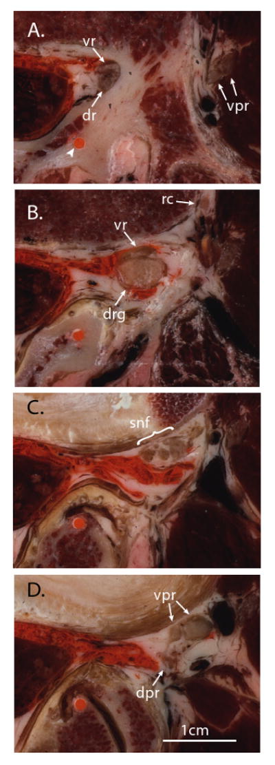
Sequential cryomicrotome sections of the adult human intervertebral foramen between the third and fourth lumbar vertebrae, demonstrating the structure of the proximal spinal nerve. The specimen was injected with orange ink by an epidural approach prior to freezing and sectioning. The orange dot (arrowhead) is an ink-filled drill hole used for guiding alignment. Medial is left in the image, anterior is up; the scale bar is common to all panels. A. At the level of the pedicle, the ventral root (vr) and dorsal root (dr) have diverged from the thecal sac in a common dural sleeve and are apposed to the medial aspect of the pedicle. The ventral primary rami (vpr) of the next rostral level are seen entering the substance of the psoas muscle. B. Somewhat more caudal, at the level of the intervertebral foramen, the ventral root and dorsal root ganglion (drg) are well outside the spinal canal, where injectate has followed. A ramus communicans (rc) is traveling posteriorly from the paravertebral sympathetic chain (off the anterior edge of the image). C. At a more caudal level, even with the rostral margin of the intervertebral disc, the dorsal root ganglion has split into fascicles that merge with similar components from the ventral root and ramus communicans to form a small plexus, composing the initial fascicles of the spinal nerve (snf). D. At a somewhat more caudal level, even with the rostral end of the intervertebral foramen, the fascicles merge into the ventral and dorsal primary rami of the spinal nerve (vpr, dpr).
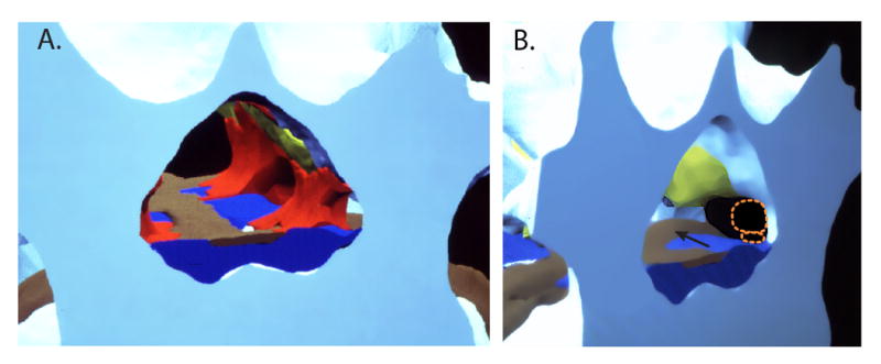
Digital reconstruction of human cryomicrotome axial images (using Skandha, copyright by the Department of Biologic Structures, University of Washington, Seattle, Washington), demonstrating the normal anatomy of the spinal canal and intervertebral foramen. A. Looking caudally from the plane of the 3rd lumbar vertebral pedicles, the medial apertures of the left L3/4 and L4/5 are apparent where they branch off from the vertebral canal. Bone is white/blue, fat is red, ligamentum flavum is yellow, the venous system is dark blue, and the disc and posterior longitudinal ligament are brown. Empty spaces would be occupied by the thecal sac in the spinal canal and the nerve root sleeve in the intervertebral foramen. Abundant fat is present in the “axilla” area between the main dural sac and the diverging nerve root sleeve. B. A more oblique view of the same digitized specimen, with the fat removed to show the structures that may impinge on nerve tissue. The dorsal root ganglion and ventral root (shown in dotted orange outline) are in the rostral end of the foramen clear of the disc, but may be compressed if loss of disc height decreases the rostro-caudal dimension of the foramen. The most common location of disc herniation is parasagittal (arrow), which most often affects the nerve roots that are heading for their exit at the next lower foramen.
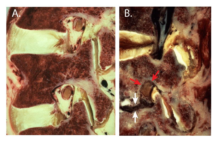
Anatomy of the intervertebral foramen in human paramedian sagittal cryomicrotome sections. A. The L3/4 and L4/5 foramina in a normal specimen are the shape of an inverted teardrop. At this lateral plane, the dorsal root ganglion is apposed to the pedicle and the ligamentum flavum. B. Disc collapse (white arrows) results in compression of the dorsal root ganglion (red arrows) through the combined effects of entry of extruded disc material (dark) into the inferior portion of the foramen, rostro-caudal shortening of the foramen, and buckling of the ligamentum flavum forward against the ganglion.
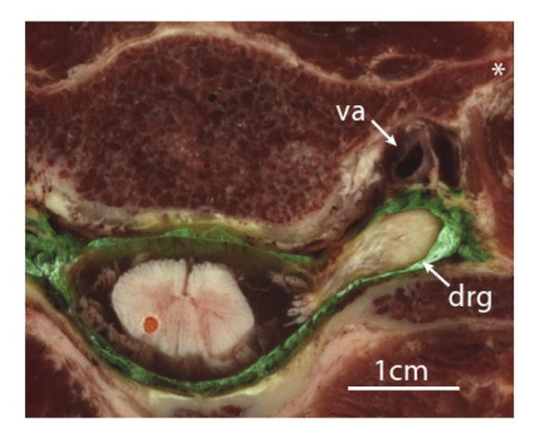
Axial cryomicrotome section through the C5/6 intervertebral foramen in a human specimen injected with green ink by an epidural approach prior to freezing and sectioning. The dorsal root ganglion (drg) lies outside the intervertebral foramen, posterior to the vertebral artery (va). The apparent discontinuity of the roots with the spinal cord is due to their sloping course that takes them out of the plane of the image. Injection into the ganglion would readily convey even a small volume of injected solution into the cord. Also shown is the anterior tubercle of the transverse process (*).
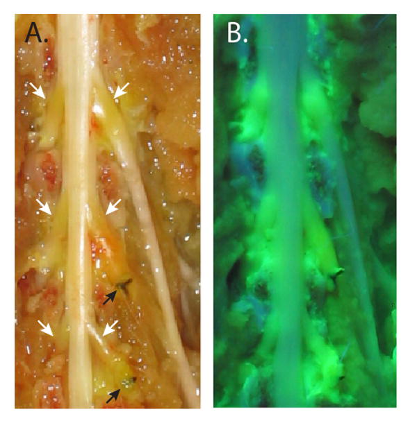
The dorsal aspect of the rat dural sac and spinal nerves, exposed by the removal of the vertebral arches, following intravenous injection of fluorescein. A. Normal illumination shows fluorescein accumulation (orange discoloration) in the areas of the dorsal root ganglia (white arrows) but not in normal nerves and cord. Ligatures on the L5 and L6 spinal nerves (black arrows) are from prior surgical preparation of the spinal nerve ligation neuropathic pain model. B. Ultraviolet illumination shows extravasated fluorescein within the dorsal root ganglia, and in spinal nerve segments just proximal to the ligatures, but not in other neural tissue.
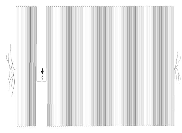
A representation of the proportionate sizes of the primary sensory neuron that would innervate the foot of an adult human, showing the soma (arrow) and central and peripheral axons (left and right). The sizes are scaled to a 50μm soma diameter, 5μm axonal diameter, and axon lengths of 30cm and 120cm. (Based on ref. 5, by permission.)

Electron microscopy image of rat dorsal root ganglion. A. The center of the image shows the soma of a sensory neuron of the small-dark category. B. An envelope of satellite glial cells, including the nucleus of one, surrounds the neuronal soma (removed from the image).
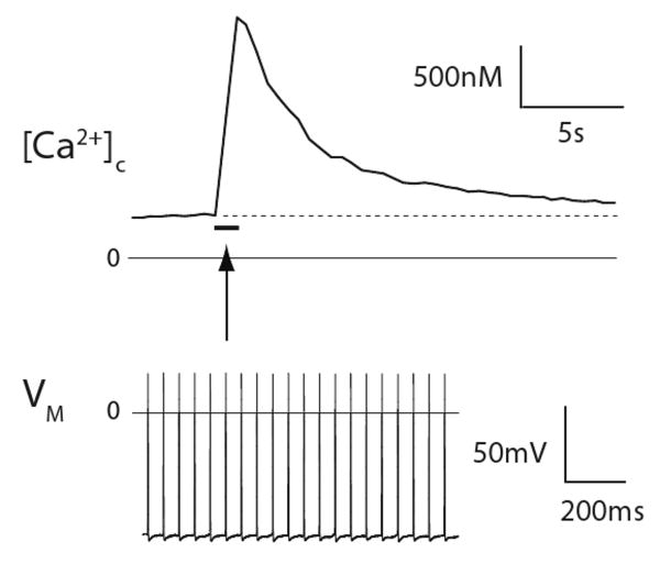
Primary sensory neuron memory, in the form of a sustained cytoplasmic Ca2+ ([Ca2+]c) signal. A 1 second train of 20 action potentials, each lasting approximately 1 millisecond (shown in the lower panel, and represented by the bar in the upper panel), produces a prolonged, 5-fold elevation of the cytoplasmic Ca2+ level (upper panel). Recordings were made simultaneously from a neuronal soma in an intact dorsal root ganglion, activated by action potentials conducted from axonal stimulation in the attached dorsal root. Dotted line in the upper panel represents resting cytoplasmic Ca2+ level. VM, transmembrane potential.
References
-
- Labat G. Regional Anesthesia: Its Technic and Clinical Application. Philadelphia: W.B. Saunders; 1922.
-
- Hogan Q. Size of human lower thoracic and lumbosacral nerve roots. Anesthesiology. 1996;85:37–42. - PubMed
-
- Kostelic J, Haughton VM, Sether L. Proximal lumbar spinal nerves in axial MR imaging, CT, and anatomic sections. Radiology. 1992;183:239–41. - PubMed
-
- Pfirrmann CW, Oberholzer PA, Zanetti M, et al. Selective nerve root blocks for the treatment of sciatica: evaluation of injection site and effectiveness--a study with patients and cadavers. Radiology. 2001;221:704–11. - PubMed
-
- Devor M. Unexplained peculiarities of the dorsal root ganglion. Pain. 1999 6:S27–35. - PubMed
Publication types
MeSH terms
Grants and funding
LinkOut - more resources
Full Text Sources