. 2010 Jul;18(7):1284-92.
doi: 10.1038/mt.2010.77. Epub 2010 May 11.

SERCA2a gene transfer enhances eNOS expression and activity in endothelial cells

Affiliations

SERCA2a gene transfer enhances eNOS expression and activity in endothelial cells

Lahouaria Hadri et al. Mol Ther. 2010 Jul.

Abstract

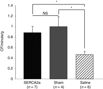
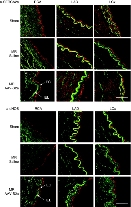
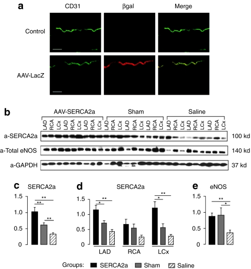
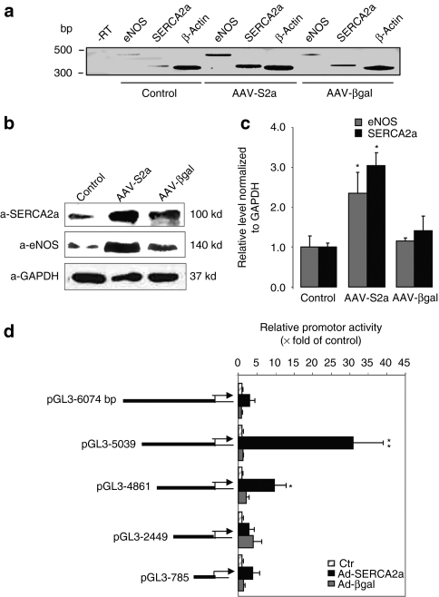
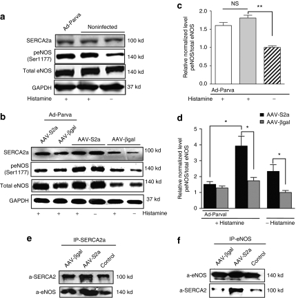
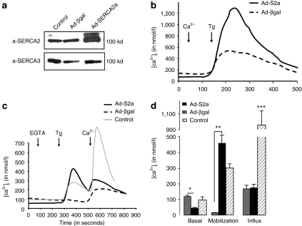
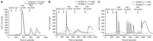
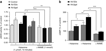
Congestive heart failure (HF) is associated with impaired endothelium-dependent nitric oxide-mediated vasodilatation. The aim of this study was to examine the effects of sarco/endoplasmic reticulum (ER) Ca(2+)-ATPase 2a (SERCA2a) gene transfer on endothelial function in a swine HF model. Two months after the creation of mitral regurgitation to induce HF, the animals underwent intracoronary injection of adeno-associated virus (AAV) carrying SERCA2a (n = 7) or saline (n = 6). At 4 months, coronary flow (CF) was measured in the mid-portion of the left anterior descending (LAD) artery. In the failing animals, CF was decreased significantly; SERCA2a gene transfer rescued CF to levels observed in sham-group [ml/min/g, 0.47 +/- 0.064 saline versus 0.89 +/- 0.116, SERCA2a; P < 0.05; 1.00 +/- 0. 185 sham P = NS (nonsignificant)]. In coronary arteries from HF animals, SERCA2a and endothelial isoform of nitric oxide synthase (eNOS) protein expression were decreased, but restored to normal levels by SERCA2a gene transfer. In human coronary artery endothelial cells (HCAECs), SERCA2a overexpression increased eNOS expression, phosphorylation, eNOS promoter activity, Ca(2+) storage capacity, and enhanced histamine-induced calcium oscillations, eNOS activity, and cyclic guanosine monophosphate (cGMP) production. Thus, SERCA2a gene transfer increases eNOS expression and activity by modulating calcium homeostasis to improve CF. These findings suggest that SERCA2a gene transfer improves vascular reactivity in the setting of HF.


Figures

Figure 1
Figure 1
SERCA2a gene transfer increases coronary blood flow in vivo in MR porcine model. Histogram showing coronary blood flow measurement in sham-operated (gray bar), MR+AAV1.SERCA2a (black bar), and MR+saline injection (hatched bars) groups. The data shown are mean ± SEM (*P < 0.05 versus saline). AAV, adeno-associated virus; MR, mitral regurgitation; NS, nonsignificant; SERCA2a, sarco/endoplasmic reticulum Ca2+-ATPase 2a.
Figure 2
Figure 2
Confocal immunofluorescence analysis of porcine coronary arteries. Cross sections of left anterior descending (LAD), left circumflex (LCx), and right coronary arteries (RCA) obtained from sham-operated or MR animals injected either with AAV.SERCA2a or with saline were analyzed by confocal immunofluorescence (red) with specific antibody; anti-SERCA2a and anti-eNOS. The media was identified by elastin autofluorescence (green). At least three animals were analyzed for each group. Bar = 50 µm. AAV, adeno-associated virus; EC, endothelial cells; eNOS, endothelial isoform of nitric oxide synthase; IEL, internal elastic lamina; M, media; MR, mitral regurgitation; SERCA2a, sarco/endoplasmic reticulum Ca2+-ATPase 2a.
Figure 3
Figure 3
SERCA2a gene transfer increases eNOS expression in coronary arteries from MR animals. (a) βgal immunostaining (red) of the left anterior descending (LAD) of a control pig (upper panel) and of a pig injected with AAV1 encoding βgal (lower panel). CD31 (green) was also used in parallel to stain EC. Bar = 50 µm. (b) Representative western blot analysis (n = 3) of SERCA2a and eNOS protein expression in coronary arteries [left circumflex (LCx), right coronary artery (RCA) and LAD] from three experimental groups: sham-operated (gray bar), MR+saline (hatched bar), and MR+AAV1.SERCA2a (black bar). (c–e) Histograms showing relative SERCA2a and eNOS ratios normalized against GAPDH used as a control. The data shown are mean ± SEM (**P < 0.01, *P < 0.05 versus saline); (c) relative expression of SERCA2a in coronary arteries from different group of animals; (d) relative expression of SERCA2a in LAD, RCA, and LCx from different animal groups; (e) relative expression of eNOS in coronary arteries from different animal groups. AAV, adeno-associated virus; βgal, β-galactosidase; EC, endothelial cell; eNOS, endothelial isoform of nitric oxide synthase; GAPDH, glyceraldehyde 3-phosphate dehydrogenase; MR, mitral regurgitation; SERCA2a, sarco/endoplasmic reticulum Ca2+-ATPase 2a.
Figure 4
Figure 4
SERCA2a overexpression increases NOS expression in HCAEC. (a) Reverse trancriptase-PCR analysis of eNOS and SERCA2a mRNAs in HCAEC cells infected with AAV1-S2a or AAV1-βgal for 6 days. β-Actin was used as an internal control. (b) Representative western blot of lysates from noninfected or infected with either AAV1-SERCA2a or AAV1-βgal HCAEC. (c) Histogram showing the relative ratio of eNOS (gray bar), and SERCA2a (black bar) normalized to GAPDH in three experiments. The values were expressed as a percentage of the value obtained for the control in the same blot. The data are mean ± SEM of three experiments (*P < 0.05 versus control). (d) Functional analysis of the human eNOS promoter in HCAEC after infection with Ad-SERCA2a or Ad-βgal. Large-scale analysis of the human eNOS promoter using deletion mutants of 6,047-bp upstream region in fusion with the luciferase gene. Relative promoter activity is expressed as a percentage of luciferase activity in control noninfected cells. Data represent the mean ± SEM of three experiments (*P < 0.05 and **P < 0.01 versus control). AAV, adeno-associated virus; Ad, adenovirus; eNOS, endothelial isoform of nitric oxide synthase; GAPDH, glyceraldehyde 3-phosphate dehydrogenase; HCAEC, human coronary artery endothelial cell; SERCA2a, sarco/endoplasmic reticulum Ca2+-ATPase 2a.
Figure 5
Figure 5
SERCA2a overexpression increases eNOS phosphorylation in Ser1177. Western blot analysis (a,b) with indicated antibodies and histogram (c,d) showing relative ratio of p(Ser1177) eNOS/total eNOS in control (c), βgal- (gray bar), or SERCA2a-infected cells (black bar) (d). The values were expressed as a percentage of the value obtained for the control or βgal nonstimulated cells in the same blot. The results are mean ± SEM of three experiments (*P < 0.05, **P < 0.01). (e,f) Immunoprecipitation analysis of functional association between SERCA2a and eNOS in HCAEC. Cell lysates were incubated with (e) SERCA2a or (f) eNOS antibodies. The immune complexes were collected with protein A/G-agarose beads and revealed by western blot analysis. eNOS, endothelial isoform of nitric oxide synthase; HCAEC, human coronary artery endothelial cell; NS, nonsignificant; SERCA2a, sarco/endoplasmic reticulum Ca2+-ATPase 2a.
Figure 6
Figure 6
SERCA2a gene transfer modulates calcium cycling in HCAEC. (a) Representative western blot of lysates from HCAEC noninfected or infected either with AAV1.SERCA2a or AAV1.βgal. Expression of SERCA2 and SERCA3 was revealed with an anti-pan SERCA2 (IID8) and an anti-pan SERCA3 (PLIM/430), respectively. (b–d) Effect of SERCA2a overexpression in HCAEC on [Ca2+]i: basal level, mobilization, and capacitative entry. At the time of the experiment (b) 1 mmol/l of CaCl2 (Ca2+) or (c) 100 µmol/l EGTA was added as indicated, and cells were then stimulated with 1 µmol/l of thapsigargin (Tg) for 4 minutes. (b) To observe the calcium capacitative entry, 300 µmol/l of CaCl2 (Ca2+) was then added to the medium. (d) Histograms showing the means ± SEM of the basal level in [Ca2+]i, Ca2+ release in response to Tg, and Ca2+ influx observed after addition of extracellular CaCl2. Representative of eight experiments obtained with four independent infections. (***P < 0.01, **P < 0.01, *P < 0.05 versus βgal). AAV1, adeno-associated virus, βgal, β-galactosidase; EGTA, ethylene glycol tetraacetic acid; HCAEC, human coronary artery endothelial cell; SERCA2a, sarco/endoplasmic reticulum Ca2+-ATPase 2a.
Figure 7
Figure 7
Effect of SERCA2a overexpression on histamine-induced calcium transient. Calcium responses with increasing histamine doses (1–10 µmol/l) in (a) control are presented as a mean of several cell (black trace) or as a single representative cell (hatched trace), and (b, c) Ad-S2a or Ad-βgal-infected HCAEC stimulated with histamine (1–10 µmol/l) in presence of extracellular calcium (100 µmol/l EGTA plus 300 µmol/l CaCl2) are presented as a mean of several cell or as a single representative cell. Ad, adenovirus; βgal, β-galactosidase; EGTA, ethylene glycol tetraacetic acid; SERCA2a, sarco/endoplasmic reticulum Ca2+-ATPase 2a; HCAEC, human coronary artery endothelial cell.
Figure 8
Figure 8
SERCA2a gene transfer increases eNOS activity and cGMP production in HCAEC. (a) Assessment of eNOS activity in HCAEC by arginine–citrulline conversion assay. Control and infected Ad-S2a or Ad-βgal cells were treated with histamine (1 µmol/l, 10 min) and pretreated with or without -NAME (1 mmol/l) for 20 minutes. The data are mean ± SEM for four experiments (*P < 0.05, **P < 0.01, ***P < 0.001 versus control). (b) cGMP production was measured using immunoassay. The values were normalized to the total protein content and were expressed as a percentage of the value obtained for the control βgal in the same experiment. The data are mean ± SEM for three experiments (*P < 0.05, **P < 0.01, ***P < 0.001 versus control). eNOS, endothelial isoform of nitric oxide synthase; HCAEC, human coronary artery endothelial cell; SERCA2a, sarco/endoplasmic reticulum Ca2+-ATPase 2a.

References

    1. Drexler H, Hayoz D, Münzel T, Just H, Zelis R., and, Brunner HR. Endothelial function in congestive heart failure. Am Heart J. 1993;126:761–764. - PubMed
    1. Kubo SH, Rector TS, Bank AJ, Williams RE., and, Heifetz SM. Endothelium-dependent vasodilation is attenuated in patients with heart failure. Circulation. 1991;84:1589–1596. - PubMed
    1. Mohri M, Egashira K, Tagawa T, Kuga T, Tagawa H, Harasawa Y, et al. Basal release of nitric oxide is decreased in the coronary circulation in patients with heart failure. Hypertension. 1997;30:50–56. - PubMed
    1. Treasure CB, Vita JA, Cox DA, Fish RD, Gordon JB, Mudge GH, et al. Endothelium-dependent dilation of the coronary microvasculature is impaired in dilated cardiomyopathy. Circulation. 1990;81:772–779. - PubMed
    1. Zhao G, Shen W, Xu X, Ochoa M, Bernstein R., and, Hintze TH. Selective impairment of vagally mediated, nitric oxide-dependent coronary vasodilation in conscious dogs after pacing-induced heart failure. Circulation. 1995;91:2655–2663. - PubMed

Publication types

MeSH terms

Substances