

Image cytometry accurately detects DNA ploidy abnormalities and predicts late relapse to high-grade dysplasia and adenocarcinoma in Barrett's oesophagus following photodynamic therapy
- PMID: 20461081
- PMCID: PMC2883155
- DOI: 10.1038/sj.bjc.6605688
Image cytometry accurately detects DNA ploidy abnormalities and predicts late relapse to high-grade dysplasia and adenocarcinoma in Barrett's oesophagus following photodynamic therapy
Abstract
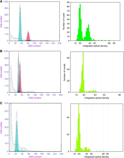
Background and aims: DNA ploidy abnormalities (aneuploidy/tetraploidy) measured by flow cytometry (FC) are strong predictors of future cancer development in untreated Barrett's oesophagus, independent of histology grade. Image cytometric DNA analysis (ICDA) is an optical technique allowing visualisation of abnormal nuclei that may be undertaken on archival tissue. Our aim was to determine the accuracy of ICDA vs FC, and evaluate DNA ploidy as a prognostic biomarker after histologically successful treatment with photodynamic therapy (PDT).
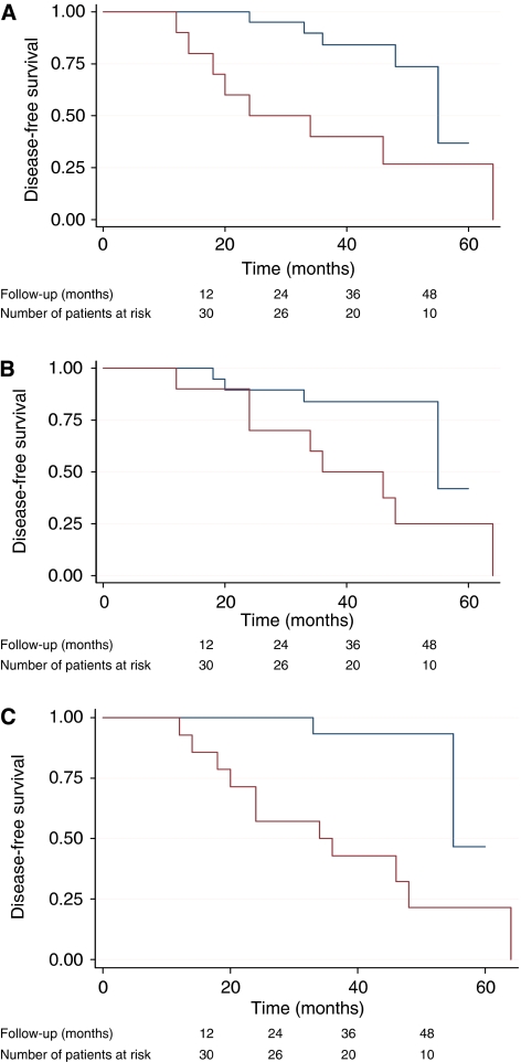
Methods: Nuclei were extracted from 40 mum sections of paraffin-embedded biopsies and processed for ICDA at UCL and FC at UW using standardised protocols. Subsequently, DNA ploidy was evaluated by ICDA on a cohort of 30 patients clear of dysplasia 1 year after aminolaevulinic acid PDT for high-grade dysplasia (HGD). The results were correlated with long-term outcome.
Results: In the comparative study, 93% (41 out of 44) of cases were classified identically. Errors occurred in the near-diploid region by ICDA and the tetraploid region by FC. In the cohort study, there were 13 cases of late relapse (7 cancer, 6 HGD) and 17 patients who remained free of dysplasia after a mean follow-up of 44 months. Aneuploidy post-PDT was highly predictive for recurrent HGD or cancer with a hazard ratio of 8.2 (1.8-37.8) (log-rank P=0.001).
Conclusions: ICDA is accurate for the detection of DNA ploidy abnormalities when compared with FC. After histologically successful PDT, patients with residual aneuploidy are significantly more likely to develop HGD or cancer than those who become diploid. DNA ploidy by ICDA is a valuable prognostic biomarker after ablative therapy.
Conflict of interest statement
The authors declare no conflict of interest.
Figures


Similar articles
-
DNA abnormalities as marker of risk for progression of Barrett's esophagus to adenocarcinoma: image cytometric DNA analysis in formalin-fixed tissues.Am J Gastroenterol. 2004 Oct;99(10):1887-94. doi: 10.1111/j.1572-0241.2004.30886.x. Am J Gastroenterol. 2004. PMID: 15447746
-
Long term efficacy of Photodynamic Therapy (PDT) as an ablative therapy of high grade dysplasia in Barrett's oesophagus.Photodiagnosis Photodyn Ther. 2013 Dec;10(4):561-5. doi: 10.1016/j.pdpdt.2013.06.002. Epub 2013 Sep 17. Photodiagnosis Photodyn Ther. 2013. PMID: 24284112 Clinical Trial.
-
Assessment of chromosomal gains as compared to DNA content changes is more useful to detect dysplasia in Barrett's esophagus brush cytology specimens.Genes Chromosomes Cancer. 2008 May;47(5):396-404. doi: 10.1002/gcc.20543. Genes Chromosomes Cancer. 2008. PMID: 18265409
-
[Flow cytometric analysis of cellular DNA content in Barret's esophagus. A study of 66 cases].Gastroenterol Clin Biol. 1991;15(10):703-10. Gastroenterol Clin Biol. 1991. PMID: 1816011 Review. French.
-
Biomarkers in Barrett's esophagus.Gastrointest Endosc Clin N Am. 2003 Apr;13(2):369-97. doi: 10.1016/s1052-5157(03)00006-0. Gastrointest Endosc Clin N Am. 2003. PMID: 12916666 Review.
Cited by
-
A biomarker panel predicts progression of Barrett's esophagus to esophageal adenocarcinoma.Dis Esophagus. 2019 Jan 1;32(1):doy102. doi: 10.1093/dote/doy102. Dis Esophagus. 2019. PMID: 30496496 Free PMC article.
-
Whole slide image cytometry: a novel method to detect abnormal DNA content in Barrett's esophagus.Lab Invest. 2015 Nov;95(11):1319-30. doi: 10.1038/labinvest.2015.98. Epub 2015 Aug 3. Lab Invest. 2015. PMID: 26237272
-
Comparison of nuclear texture analysis and image cytometric DNA analysis for the assessment of dysplasia in Barrett's oesophagus.Br J Cancer. 2011 Oct 11;105(8):1218-23. doi: 10.1038/bjc.2011.353. Epub 2011 Sep 20. Br J Cancer. 2011. PMID: 21934680 Free PMC article.
-
Robust microbial cell segmentation by optical-phase thresholding with minimal processing requirements.Cytometry A. 2017 May;91(5):443-449. doi: 10.1002/cyto.a.23099. Epub 2017 Mar 30. Cytometry A. 2017. PMID: 28371011 Free PMC article.
-
Aneuploidy in targeted endoscopic biopsies outperforms other tissue biomarkers in the prediction of histologic progression of Barrett's oesophagus: A multi-centre prospective cohort study.EBioMedicine. 2020 Jun;56:102765. doi: 10.1016/j.ebiom.2020.102765. Epub 2020 May 24. EBioMedicine. 2020. PMID: 32460165 Free PMC article.
References
-
- Baldetorp B, Ferno M, Fallenius A, Fallenius-Vecchi G, Idvall I, Olsson H, Sigurdsson H, Akerman M, Killander D (1992) Image cytometric DNA analysis in human breast cancer analysis may add prognostic information in diploid cases with low S-phase fraction by flow cytometry. Cytometry 13: 577–585 - PubMed
-
- Bocking A, Giroud F, Reith A (1995) ESACP DNA consensus in image cytometry. ACP 8: 67–74 - PubMed
-
- Buttar NS, Wang KK, Sebo TJ, Riehle DM, Krishnadath KK, Lutzke LS, Anderson MA, Petterson TM, Burgart LJ (2001) Extent of high-grade dysplasia in Barrett's esophagus correlates with risk of adenocarcinoma. Gastroenterology 120: 1630–1639 - PubMed
-
- Cameron AJ, Ott BJ, Payne WS (1985) The incidence of adenocarcinoma in columnar-lined (Barrett's) esophagus. N Engl J Med 313: 857–859 - PubMed
-
- Chen TL, Luo I, Mikhail N, Raskova J, Raska K (1995) Comparison of flow and image cytometry for Dna content-analysis of fresh and formalin-fixed, paraffin-embedded tissue in breast-carcinoma. Cytometry 22: 181–189 - PubMed
Publication types
MeSH terms
Substances
Grants and funding
LinkOut - more resources
Full Text Sources
Other Literature Sources
