

H2AX phosphorylation screen of cells from radiosensitive cancer patients reveals a novel DNA double-strand break repair cellular phenotype
- PMID: 20461094
- PMCID: PMC2869166
- DOI: 10.1038/sj.bjc.6605666
H2AX phosphorylation screen of cells from radiosensitive cancer patients reveals a novel DNA double-strand break repair cellular phenotype
Abstract
Background: About 1-5% of cancer patients suffer from significant normal tissue reactions as a result of radiotherapy (RT). It is not possible at this time to predict how most patients' normal tissues will respond to RT. DNA repair dysfunction is implicated in sensitivity to RT particularly in genes that mediate the repair of DNA double-strand breaks (DSBs). Phosphorylation of histone H2AX (phosphorylated molecules are known as gammaH2AX) occurs rapidly in response to DNA DSBs, and, among its other roles, contributes to repair protein recruitment to these damaged sites. Mammalian cell lines have also been crucial in facilitating the successful cloning of many DNA DSB repair genes; yet, very few mutant cell lines exist for non-syndromic clinical radiosensitivity (RS).
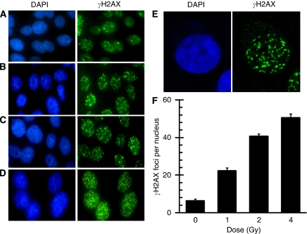
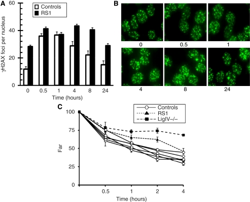
Methods: Here, we survey DNA DSB induction and repair in whole cells from RS patients, as revealed by gammaH2AX foci assays, as potential predictive markers of clinical radiation response.
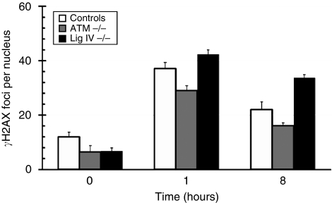
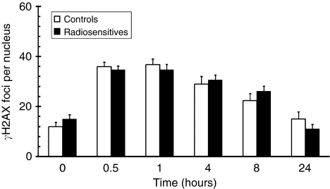
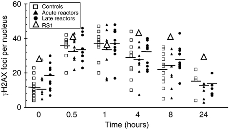
Results: With one exception, both DNA focus induction and repair in cell lines from RS patients were comparable with controls. Using gammaH2AX foci assays, we identified a RS cancer patient cell line with a novel ionising radiation-induced DNA DSB repair defect; these data were confirmed by an independent DNA DSB repair assay.
Conclusion: gammaH2AX focus measurement has limited scope as a pre-RT predictive assay in lymphoblast cell lines from RT patients; however, the assay can successfully identify novel DNA DSB repair-defective patient cell lines, thus potentially facilitating the discovery of novel constitutional contributions to clinical RS.
Figures
Similar articles
-
Early increase of radiation-induced γH2AX foci in a human Ku70/80 knockdown cell line characterized by an enhanced radiosensitivity.J Radiat Res. 2010;51(6):633-41. doi: 10.1269/jrr.10033. J Radiat Res. 2010. PMID: 21116096
-
DNA double-strand break repair of blood lymphocytes and normal tissues analysed in a preclinical mouse model: implications for radiosensitivity testing.Clin Cancer Res. 2008 Oct 15;14(20):6546-55. doi: 10.1158/1078-0432.CCR-07-5147. Clin Cancer Res. 2008. PMID: 18927295
-
DNA repair alterations in children with pediatric malignancies: novel opportunities to identify patients at risk for high-grade toxicities.Int J Radiat Oncol Biol Phys. 2010 Oct 1;78(2):359-69. doi: 10.1016/j.ijrobp.2009.08.052. Epub 2010 Feb 12. Int J Radiat Oncol Biol Phys. 2010. PMID: 20153123
-
Ability to repair DNA double-strand breaks related to cancer susceptibility and radiosensitivity.Radiat Med. 2007 Nov;25(9):433-8. doi: 10.1007/s11604-007-0161-3. Epub 2007 Nov 26. Radiat Med. 2007. PMID: 18026900 Review.
-
Mechanism of elimination of phosphorylated histone H2AX from chromatin after repair of DNA double-strand breaks.Mutat Res. 2010 Mar 1;685(1-2):54-60. doi: 10.1016/j.mrfmmm.2009.08.001. Epub 2009 Aug 12. Mutat Res. 2010. PMID: 19682466 Review.
Cited by
-
In vitro prediction of breast cancer therapy toxicity.Ann Transl Med. 2017 Mar;5(5):94. doi: 10.21037/atm.2017.02.27. Ann Transl Med. 2017. PMID: 28361059 Free PMC article.
-
Changing of gamma-H2AX in peripheral blood mononuclear cells during concurrent chemoradiation in locally advanced rectal cancer patients: a potential response predictor.J Gastrointest Oncol. 2024 Oct 31;15(5):2117-2128. doi: 10.21037/jgo-24-488. Epub 2024 Oct 29. J Gastrointest Oncol. 2024. PMID: 39554568 Free PMC article.
-
Shifts in developmental timing, and not increased levels of experience-dependent neuronal activity, promote barrel expansion in the primary somatosensory cortex of rats enucleated at birth.PLoS One. 2013;8(1):e54940. doi: 10.1371/journal.pone.0054940. Epub 2013 Jan 25. PLoS One. 2013. PMID: 23372796 Free PMC article.
-
ZSCAN4 interacts with PARP1 to promote DNA repair in mouse embryonic stem cells.Cell Biosci. 2023 Oct 24;13(1):193. doi: 10.1186/s13578-023-01140-1. Cell Biosci. 2023. PMID: 37875990 Free PMC article.
-
The ascidian natural product eusynstyelamide B is a novel topoisomerase II poison that induces DNA damage and growth arrest in prostate and breast cancer cells.Oncotarget. 2015 Dec 22;6(41):43944-63. doi: 10.18632/oncotarget.6267. Oncotarget. 2015. PMID: 26733491 Free PMC article.
References
-
- Alsner J, Andreassen CN, Overgaard J (2008) Genetic markers for prediction of normal tissue toxicity after radiotherapy. Semin Radiat Oncol 18: 126–135 - PubMed
-
- Banath JP, Macphail SH, Olive PL (2004) Radiation sensitivity, H2AX phosphorylation, and kinetics of repair of DNA strand breaks in irradiated cervical cancer cell lines. Cancer Res 64: 7144–7149 - PubMed
-
- Bassing CH, Alt FW (2004) H2AX may function as an anchor to hold broken chromosomal DNA ends in close proximity. Cell Cycle 3: 149–153 - PubMed
-
- Bassing CH, Chua KF, Sekiguchi J, Suh H, Whitlow SR, Fleming JC, Monroe BC, Ciccone DN, Yan C, Vlasakova K, Livingston DM, Ferguson DO, Scully R, Alt FW (2002) Increased ionizing radiation sensitivity and genomic instability in the absence of histone H2AX. Proc Natl Acad Sci USA 99: 8173–8178 - PMC - PubMed
-
- Bouquet F, Muller C, Salles B (2006) The loss of gammaH2AX signal is a marker of DNA double strand breaks repair only at low levels of DNA damage. Cell Cycle 5: 1116–1122 - PubMed
Publication types
MeSH terms
Substances
LinkOut - more resources
Full Text Sources
Other Literature Sources
