. 2010 Oct;18(10):1133-40.
doi: 10.1038/ejhg.2010.59. Epub 2010 May 12.

High frequency of COH1 intragenic deletions and duplications detected by MLPA in patients with Cohen syndrome

Affiliations

High frequency of COH1 intragenic deletions and duplications detected by MLPA in patients with Cohen syndrome

Veronica Parri et al. Eur J Hum Genet. 2010 Oct.

Abstract

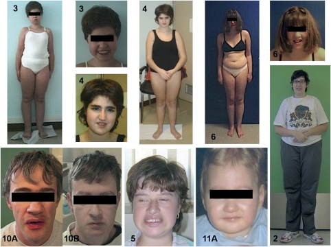
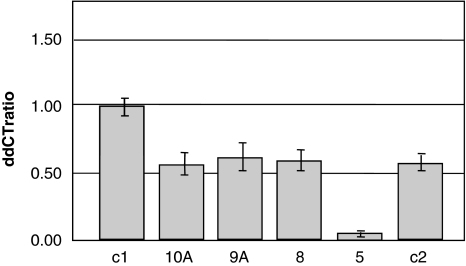
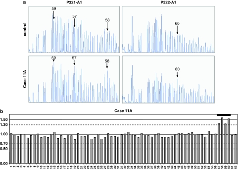
Cohen syndrome is a rare, clinically variable autosomal recessive disorder characterized by mental retardation, postnatal microcephaly, facial dysmorphisms, ocular abnormalities and intermittent neutropenia. Mutations in the COH1 gene have been found in patients from different ethnic origins. However, a high percentage of patients have only one or no mutated allele. To investigate whether COH1 copy number changes account for missed mutations, we used multiplex ligation-dependent probe amplification (MLPA) to test a group of 14 patients with Cohen syndrome. This analysis has allowed us to identify multi-exonic deletions in 11 alleles and duplications in 4 alleles. Considering our previous study, COH1 copy number variations represent 42% of total mutated alleles. To our knowledge, COH1 intragenic duplications have never been reported in Cohen syndrome. The three duplications encompassed exons 4-13, 20-30 and 57-60, respectively. Interestingly, four deletions showed the same exon coverage (exons 6-16) with respect to a deletion recently reported in a large Greek consanguineous family. Haplotype analysis suggested a possible founder effect in the Mediterranean basin. The use of MLPA was therefore crucial in identifying mutated alleles undetected by traditional techniques and in defining the extent of the deletions/duplications. Given the high percentage of identified copy number variations, we suggest that this technique could be used as the initial screening method for molecular diagnosis of Cohen syndrome.


Figures

Figure 1
Figure 1
Clinical features of Cohen syndrome patients. Note the typical facial gestalt of patients 3, 4, 5, 6, 10A, 10B and 11A. Frontal views of patients 2, 3, 4 and 6, showing truncal obesity.
Figure 2
Figure 2
MLPA analysis results showing the recurrent deletion in heterozygous (Case 8) and homozygous (Case 5) states. (a) Electropherograms obtained with P321-A1 kit (on the left) and P322-A1 kit (on the right) for a normal control sample, patient 8 and patient 5. Numbers and arrows indicate the exon probes with reduced fluorescence signals with respect to the control sample. In patient 8, the signal is half-reduced for probes 7–16, whereas in patient 5 there is no signal for the same probes. (b, c) Peak area histograms for patients 8 (a) and 5 (b) normalized with the control sample. Exon dosage is reported on the y axis (normal values spanning from 0.7 to 1.3 are indicated with broken lines). MLPA analysis shows reduced peak area for exons 7–16, compatible with a heterozygous deletion in patient 8 and a homozygous deletion in patient 5. Deletions are indicated with a heavy black line.
Figure 3
Figure 3
Quantitative PCR results for exon 6 of COH1. ddCT ratios and standard deviations of a normal control sample (c1), a deleted control sample (c2) and patients 5, 8, 9A and 10A. Compared with controls, patients 8, 9A and 10A show ddCT ratio values of about 0.5, indicating a deletion in the heterozygous state, whereas patient 5 shows ddCT ratio values of about 0.0, indicating a deletion in the homozygous state.
Figure 4
Figure 4
MLPA analysis results showing the duplication spanning exons 57–60 in the familial case with an affected brother (11A) and sister (11B). (a) Electropherograms obtained with P321-A1 kit (on the left) and P322-A1 kit (on the right) for a normal control sample and patient 11A. Numbers and arrows indicate the exon probes with increased fluorescence signals with respect to the control sample. (b) Peak area histograms for patient 11A normalized with the control sample. The exon dosage is reported on the y axis (normal values spanning from 0.7 to 1.3 are indicated with broken lines). The consistent increase in the peak area for exons 57–60 is compatible with a duplication of these exons (indicated with a heavy black line).
Figure 5
Figure 5
Characterization of duplication 57–60 in familial case 11. (a) Schematic drawing of the duplicated region. The star indicates the position of the MLPA probe in exon 61, whereas the thunder represents the insertion point of the duplicated segment. Arrows indicate the primers located within introns 59 and 56 used in the long-range PCR experiment. (b) Sequence analysis showing the junction between intron 56 and exon 61. (c) Aligned exon 61 and intron 56 sequences at the duplication junction. Region of homology across the duplication junction is boxed.

Similar articles

Cited by

References

    1. Cohen MM, Jr, Hall BD, Smith DW, Graham CB, Lampert KJ. A new syndrome with hypotonia, obesity, mental deficiency, and facial, oral, ocular, and limb anomalies. J Pediatr. 1973;83:280–284. - PubMed
    1. Carey JC, Hall BD. Confirmation of the Cohen syndrome. J Pediatr. 1978;93:239–244. - PubMed
    1. Norio R, Raitta C, Lindahl E. Further delineation of the Cohen syndrome; report on chorioretinal dystrophy, leukopenia and consanguinity. Clin Genet. 1984;25:1–14. - PubMed
    1. Kolehmainen J, Black GC, Saarinen A, et al. Cohen syndrome is caused by mutations in a novel gene, COH1, encoding a transmembrane protein with a presumed role in vesicle-mediated sorting and intracellular protein transport. Am J Hum Genet. 2003;72:1359–1369. - PMC - PubMed
    1. Velayos-Baeza A, Vettori A, Copley RR, Dobson-Stone C, Monaco AP. Analysis of the human VPS13 gene family. Genomics. 2004;84:536–549. - PubMed

MeSH terms

Substances

Supplementary concepts