

Case Reports
doi: 10.3340/jkns.2010.47.4.306.
Epub 2010 Apr 30.
Synovial sarcoma of the posterior neck : a case report and review of literature
Affiliations
- PMID: 20461175
- PMCID: PMC2864827
- DOI: 10.3340/jkns.2010.47.4.306
Item in Clipboard
Case Reports
Synovial sarcoma of the posterior neck : a case report and review of literature
J Korean Neurosurg Soc.
2010 Apr.
Abstract
We recently experienced a case of synovial sarcoma in the posterior neck, which involved adjacent bony structures. Synovial sarcoma is rare, malignant soft tissue tumor that occur predominantly in the lower extremities. Wide surgical excision with involved tissue is the treatment of first choice, because most synovial sarcomas reveal aggressive features. We removed the tumor with involved bony structures and patient was given postoperative radiation therapy. Despite these treatment options, the patient died 1 year after surgery. We report this case with a review of the literature.
Keywords: Bony involvement; Posterior neck; Synovial sarcoma.
Figures
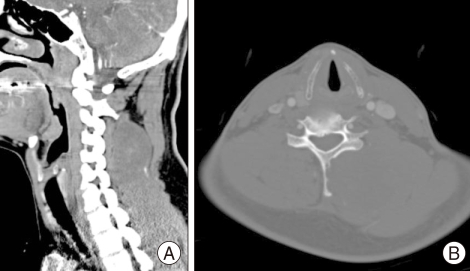
A : Non-enhanced neck computed tomography (CT) scan shows a mass lesion with mixed density in the left paraspinal area. B : Neck CT scan in bone setting showed destruction of the spinous process and lamina of cervical vertebra.
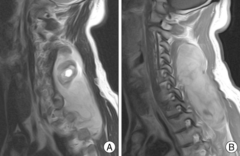
A : Cervical T2-weighted sagittal magnetic resonance (MR) image reveals mixed signal (mainly high signal intensity) in the lesion with an internal cystic component. B : Cervical T1-weighted sagittal enhanced MR image showing the heterogenous enhanced, well demarcated mass lesion in the posterior neck.
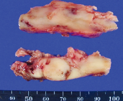
In gross finding, the removed mass is well capsulated, soft, gray colored, and low vascular lesions. The upper part of the tumor consists of old hematoma.
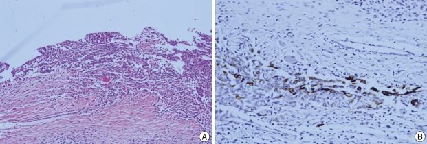
A : Microscopically, the tumor consists of spindle cell and epithelial cell components. B : On immunohistochemistry, the tumor isfocally stained by CK-7.
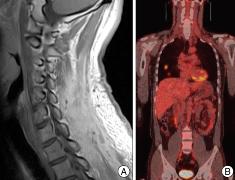
A : Six months after surgery, a follow up MR image shows no recurrent lesion on the previous operation site. B : Follow up positron emission tomography (PET) scan reveals multiple lung metastasis.
Similar articles
-
Synovial sarcoma of the head and neck: a review of its diagnosis and management and a report of a rare case of orbital involvement.Ear Nose Throat J. 2010 Jun;89(6):280-3. Ear Nose Throat J. 2010. PMID: 20556741 Review.
-
Synovial Sarcoma of Cheek: A Rare Case Report with Review of Literature.Indian J Otolaryngol Head Neck Surg. 2023 Jun;75(2):913-916. doi: 10.1007/s12070-022-03189-9. Epub 2022 Oct 3. Indian J Otolaryngol Head Neck Surg. 2023. PMID: 37275007 Free PMC article.
-
A rare intraosseous synovial sarcoma of the mandible: A case report.Int J Surg Case Rep. 2024 Jul;120:109880. doi: 10.1016/j.ijscr.2024.109880. Epub 2024 Jun 11. Int J Surg Case Rep. 2024. PMID: 38870657 Free PMC article.
-
Biphasic synovial sarcoma of the epiglottis: Case report and literature review.Auris Nasus Larynx. 2018 Jun;45(3):617-621. doi: 10.1016/j.anl.2017.06.007. Epub 2017 Jul 6. Auris Nasus Larynx. 2018. PMID: 28689931 Review.
-
Synovial sarcoma of the maxillary sinus: an extremely rare case with excellent response to chemotherapy.Onco Targets Ther. 2018 Jan 23;11:483-488. doi: 10.2147/OTT.S151473. eCollection 2018. Onco Targets Ther. 2018. PMID: 29416348 Free PMC article.
Cited by
-
Do Posterior Neck Lumps Need Ultrasound Evaluation: A Case Series of 623 Neck Ultrasound Studies at a Single Institution.J Prim Care Community Health. 2024 Jan-Dec;15:21501319241271284. doi: 10.1177/21501319241271284. J Prim Care Community Health. 2024. PMID: 39105339 Free PMC article.
-
Clinical, pathological and unusual MRI features of five synovial sarcomas in head and neck.Br J Radiol. 2015 Jun;88(1050):20140843. doi: 10.1259/bjr.20140843. Br J Radiol. 2015. PMID: 25945512 Free PMC article.
-
Tension Pneumothorax: Is it Sarcoma or Pazopanib?Cureus. 2020 Oct 14;12(10):e10945. doi: 10.7759/cureus.10945. Cureus. 2020. PMID: 33200058 Free PMC article.
-
Primary giant mediastinal synovial sarcoma of the neck: A case report and review of the literature.Oncol Lett. 2014 Jan;7(1):140-144. doi: 10.3892/ol.2013.1649. Epub 2013 Oct 29. Oncol Lett. 2014. PMID: 24348836 Free PMC article.
-
A Rare Case of Synovial Sarcoma Involving the Brachial Plexus, Treated with Wide Local Excision and Reconstructed with Sural Nerve Grafts.Indian J Surg Oncol. 2019 Sep;10(3):435-436. doi: 10.1007/s13193-019-00883-z. Epub 2020 Aug 14. Indian J Surg Oncol. 2019. PMID: 32831528 Free PMC article.
References
-
- Alektiar KM, Leung D, Zelefsky MJ, Brennan MF. Adjuvant radiation for stage II-B soft tissue sarcoma of the extremity. J Clin Oncol. 2002;20:1643–1650. - PubMed
-
- Bergh P, Meis-Kindblom JM, Gherlinzoni F, Berlin O, Bacchini P, Bertoni F, et al. Synovial sarcoma : identification of low and high risk groups. Cancer. 1999;85:2596–2607. - PubMed
-
- Davis AM, O'Sullivan B, Turcotte R, Bell R, Catton C, Chabot P, et al. Late radiation morbidity following randomization to preoperative versus postoperative radiotherapy in extremity soft tissue sarcoma. Radiother Oncol. 2005;75:48–53. - PubMed
Publication types
LinkOut - more resources
Full Text Sources
