. 2010 May-Jun;11(3):295-303.
doi: 10.3348/kjr.2010.11.3.295. Epub 2010 Apr 29.

Apparent diffusion coefficient value of diffusion-weighted imaging for hepatocellular carcinoma: correlation with the histologic differentiation and the expression of vascular endothelial growth factor

Affiliations

Apparent diffusion coefficient value of diffusion-weighted imaging for hepatocellular carcinoma: correlation with the histologic differentiation and the expression of vascular endothelial growth factor

Suk Hee Heo et al. Korean J Radiol. 2010 May-Jun.

Abstract

Objective: To evaluate whether the histopathological differentiation and the expression of vascular endothelial growth factor (VEGF) of hepatocellular carcinoma (HCC) do show correlation with the apparent diffusion coefficient (ADC) value on diffusion-weighted imaging (DWI).

Materials and methods: Twenty-seven HCCs from 27 patients who had undergone preoperative liver MRI (1.5T) and surgical resection were retrospectively reviewed. DWI was obtained with a single-shot, echo-planar imaging sequence in the axial plane (b values: 0 and 1,000 sec/mm(2)). On DWIs, the ADC value of the HCCs was measured by one radiologist, who was kept 'blinded' to the histological findings. Histopathologically, the differentiation was classified into well (n = 9), moderate (n = 9) and poor (n = 9). The expression of VEGF was semiquantitatively graded as grade 0 (n = 8), grade 1 (n = 9) and grade 2 (n = 10). We analyzed whether the histopathological differentiation and the expression of VEGF of the HCC showed correlation with the ADC value on DWI.

Results: The mean ADC value of the poorly-differentiated HCCs (0.9 +/- 0.13x10(-3) mm(2)/s) was lower than those of the well-differentiated HCCs (1.2 +/- 0.22x10(-3) mm(2)/s) (p = 0.031) and moderately-differentiated HCCs (1.1 +/- 0.01x10(-3) mm(2)/s) (p = 0.013). There was a significant correlation between the differentiation and the ADC value of the HCCs (r = -0.51, p = 0.012). The mean ADC of the HCCs with a VEGF expression grade of 0, 1 and 2 was 1.1 +/- 0.17, 1.1 +/- 0.21 and 1.1 +/- 0.18x10(-3) mm(2)/s, respectively. The VEGF expression did not show correlation with the ADC value of the HCCs (r = 0.07, p = 0.74).

Conclusion: The histopathological differentiation of HCC shows inverse correlation with the ADC value. Therefore, DWI with ADC measurement may be a valuable tool for noninvasively predicting the differentiation of HCC.

Keywords: Liver neoplasm; Liver neoplasm, MR; Magnetic resonance (MR), diffusion study.


Figures

Fig. 1
Fig. 1
Method of apparent diffusion coefficient measurement. Apparent diffusion coefficient values were obtained by using five ROIs with uniform size (42 pixels) on apparent diffusion coefficient map (A), placed on area corresponding to enhancing solid portion of HCCs demonstrated on contrast-enhanced T1-weighted image (B). ROIs were carefully placed on solid portion to avoid cystic or necrotic portion.
Fig. 2
Fig. 2
Graphs of apparent diffusion coefficient value (×10-3 mm2/s) of hepatocellular carcinomas based on differentiation and vascular endothelial growth factor expression. Outlier indicates range; from largest to smallest observed data points within 1.5 interquartile range presented by box. Horizontal line is median (50th percentile) of measured values; top and bottom of box represent 25th and 75th percentiles, respectively. ADC = apparent diffusion coefficient, VEGF = vascular endothelial growth factor A. There was significant difference in apparent diffusion coefficient values among well-, moderately- and poorly-differentiated hepatocellular carcinomas (p = 0.026, Kruskal-Wallis test). For pair-wise comparisons, apparent diffusion coefficient value of poorly-differentiated hepatocellular carcinomas was significantly lower than that of moderately-differentiated hepatocellular carcinomas (p = 0.013). B. There was no significant difference between vascular endothelial growth factor expression and apparent diffusion coefficient value for hepatocellular carcinomas.
Fig. 3
Fig. 3
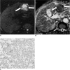
51-year-old man with poorly-differentiated hepatocellular carcinoma. A. Axial diffusion-weighted image (TR/TE, 8,000 ms/92 ms; and b - 1,000 sec/mm2) shows 4 × 3.5 cm hepatocellular carcinoma with high signal intensity in left hepatic lobe (arrows). B. On diffusion coefficient map, apparent diffusion coefficient value of tumor (arrows) was 0.88 ± 0.04 (mean ± SD, ×10-3 mm2/s). C. Photomicrograph (Hematoxylin & Eosin staining, ×200) reveals poorly-differentiated hepatocellular carcinoma with irregular nuclei, marked hyperchromatism, and increased mitoses.
Fig. 4
Fig. 4
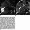
63-year-old man with moderately-differentiated hepatocellular carcinoma. A. Axial diffusion-weighted image (TR/TE, 8,000 ms/92 ms; and b - 1,000 sec/mm2) shows 5 × 3 cm hepatocellular carcinoma with high signal intensity in right hepatic lobe (arrows). B. On diffusion coefficient map, apparent diffusion coefficient value of tumor (arrows) was 1.14 ± 0.04 (mean ± SD, ×10-3 mm2/s). C. Photomicrograph (Hematoxylin & Eosin staining, × 200) demonstrates moderately-differentiated hepatocellular carcinoma with round nuclei and relative decrease in mitotic activity.

References

    1. Ince N, Wands JR. The increasing incidence of hepatocellular carcinoma. N Engl J Med. 1999;340:798–799. - PubMed
    1. Tung-Ping Poon R, Fan ST, Wong J. Risk factors, prevention, and management of postoperative recurrence after resection of hepatocellular carcinoma. Ann Surg. 2000;232:10–24. - PMC - PubMed
    1. Haratake J, Takeda S, Kasai T, Nakano S, Tokui N. Predictable factors for estimating prognosis of patients after resection of hepatocellular carcinoma. Cancer. 1993;72:1178–1183. - PubMed
    1. Kim SH, Lee WJ, Lim HK, Park CK. SPIO-enhanced MRI findings of well-differentiated hepatocellular carcinomas: correlation with MDCT findings. Korean J Radiol. 2009;10:112–120. - PMC - PubMed
    1. Yamaguchi R, Yano H, Iemura A, Ogasawara S, Haramaki M, Kojiro M. Expression of vascular endothelial growth factor in human hepatocellular carcinoma. Hepatology. 1998;28:68–77. - PubMed

Publication types

MeSH terms