

Mesothelial cyst of the round ligament mimicking a metastasis: a case report
- PMID: 20461192
- PMCID: PMC2864865
- DOI: 10.3348/kjr.2010.11.3.364
Mesothelial cyst of the round ligament mimicking a metastasis: a case report
Abstract
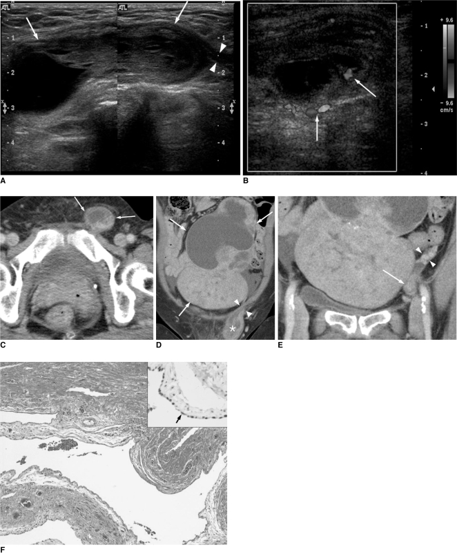
A mesothelial cyst of the round ligament is a rare cause of an inguinal mass. Clinically, it is frequently misdiagnosed as one of commoner diseases such as an inguinal hernia, femoral hernia, lipoma, and lymphadenopathy upon physical examination. Some previous reports elaborated the sonographic features of a mesothelial cyst of the round ligament. However, to our knowledge, few reports have described the CT features of a mesothelial cyst. We illustrated here the sonographic and multidetector CT features of a case of a mesothelial cyst of the round ligament that presented as an inguinal palpable mass and mimicked a metastasis in a patient with a Sertoli-Leydig cell tumor of the ovary.
Keywords: Inguinal area; Mesothelial cyst; Multi-detector row CT; Round ligament; Ultrasonography.
Figures
References
-
- Harper GB, Jr, Awbrey BJ, Thomas CG, Jr, Askin FB. Mesothelial cysts of the round ligament simulating inguinal hernia. Report of four cases and a review of the literature. Am J Surg. 1986;151:515–517. - PubMed
-
- Oh SN, Jung SE, Rha SE, Lim GY, Ku YM, Byun JY, et al. Sonography of various cystic masses of the female groin. J Ultrasound Med. 2007;26:1735–1742. - PubMed
-
- Warshauer DM, Mandel SR. Leiomyoma of the extraperitoneal round ligament: CT demonstration. Clin Imaging. 1999;23:375–376. - PubMed
-
- Oh SN, Jung SE, Lee JM, Chung JH, Park GS. Sonographic diagnosis of a round ligament cyst in the inguinal area. J Clin Ultrasound. 2007;35:226–228. - PubMed
-
- Khanna PC, Ponsky T, Zagol B, Lukish JR, Markle BM. Sonographic appearance of canal of Nuck hydrocele. Pediatr Radiol. 2007;37:603–606. - PubMed
Publication types
MeSH terms
Substances
LinkOut - more resources
Full Text Sources
Medical
