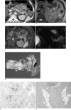
Imaging findings in a case of mixed acinar-endocrine carcinoma of the pancreas
- PMID: 20461195
- PMCID: PMC2864868
- DOI: 10.3348/kjr.2010.11.3.378
Imaging findings in a case of mixed acinar-endocrine carcinoma of the pancreas
Abstract
Mixed acinar-endocrine carcinoma (MAEC) of the pancreas is extremely uncommon. We report here a rare case of MAEC of the pancreas presenting as watery diarrhea. This is the first report in the English-language literature that describes the imaging findings of MAEC of the pancreas, including computed tomography (CT), magnetic resonance (MR) imaging, and MR cholangiopancreatography features.
Keywords: Endocrine; Exocrine; Mixed acinar-endocrine carcinoma; Pancreas.
Figures

Similar articles
-
Intraductal growing acinar cell carcinoma of the pancreas.Abdom Imaging. 2013 Oct;38(5):1115-9. doi: 10.1007/s00261-013-9993-8. Abdom Imaging. 2013. PMID: 23515859
-
Uncommon solid pancreatic neoplasms: ultrasound, computed tomography, and magnetic resonance imaging features.Semin Ultrasound CT MR. 2007 Oct;28(5):357-70. doi: 10.1053/j.sult.2007.08.002. Semin Ultrasound CT MR. 2007. PMID: 17970552 Review.
-
Mixed acinar-endocrine carcinoma of the pancreas with intraductal growth into the main pancreatic duct: Report of a case.Surg Today. 2010 Apr;40(4):380-4. doi: 10.1007/s00595-009-4083-9. Epub 2010 Mar 26. Surg Today. 2010. PMID: 20339996
-
Omental acinar cell carcinoma of pancreatic origin in a child: a clinicopathological rarity.Pediatr Surg Int. 2016 Mar;32(3):307-11. doi: 10.1007/s00383-015-3850-5. Epub 2015 Dec 22. Pediatr Surg Int. 2016. PMID: 26694824
-
[PET and PET-CT of malignant tumors of the exocrine pancreas].Radiologe. 2009 Feb;49(2):131-6. doi: 10.1007/s00117-008-1756-0. Radiologe. 2009. PMID: 19219476 Review. German.
Cited by
-
Mixed Acinar-Endocrine Carcinoma (MAEC) Arising in Duodenal Pancreatic Heterotopia.Case Rep Pathol. 2019 Sep 2;2019:1713546. doi: 10.1155/2019/1713546. eCollection 2019. Case Rep Pathol. 2019. PMID: 31565458 Free PMC article.
-
Peri-ampullary mixed acinar-endocrine carcinoma.Rare Tumors. 2011 Apr 4;3(2):e15. doi: 10.4081/rt.2011.e15. Rare Tumors. 2011. PMID: 21769314 Free PMC article.
-
Mixed acinar-endocrine carcinoma of the pancreas treated with S-1.Clin J Gastroenterol. 2013 Dec;6(6):459-64. doi: 10.1007/s12328-013-0416-8. Epub 2013 Sep 5. Clin J Gastroenterol. 2013. PMID: 26182137
-
Mixed acinar-endocrine carcinoma of pancreas: A case report and brief review of the literature.Onco Targets Ther. 2015 Jul 3;8:1633-42. doi: 10.2147/OTT.S87406. eCollection 2015. Onco Targets Ther. 2015. PMID: 26170699 Free PMC article.
-
Mixed adeno(neuro)endocrine carcinoma arising from the ectopic gastric mucosa of the upper thoracic esophagus.World J Surg Oncol. 2013 Sep 4;11:218. doi: 10.1186/1477-7819-11-218. World J Surg Oncol. 2013. PMID: 24139488 Free PMC article.
References
-
- Kamisawa T, Tu Y, Egawa N, Ishiwata J, Tsuruta K, Okamoto A, et al. Ductal and acinar differentiation in pancreatic endocrine tumors. Dig Dis Sci. 2002;47:2254–2261. - PubMed
-
- Klöppel G. Mixed exocrine-endocrine tumors of the pancreas. Semin Diagn Pathol. 2000;17:104–108. - PubMed
-
- Schron DS, Mendelsohn G. Pancreatic carcinoma with duct, endocrine, and acinar differentiation. A histologic, immunocytochemical, and ultrastructural study. Cancer. 1984;54:1766–1770. - PubMed
-
- Ohike N, Kosmahl M, Klöppel G. Mixed acinar-endocrine carcinoma of the pancreas. A clinicopathological study and comparison with acinar-cell carcinoma. Virchows Arch. 2004;445:231–235. - PubMed
-
- Cubilla AL, Fitzgerald PJ. Cancer of the exocrine pancreas: the pathologic aspects. CA Cancer J Clin. 1985;35:2–18. - PubMed
Publication types
MeSH terms
LinkOut - more resources
Full Text Sources
Medical

