C5 palsy following anterior decompression and spinal fusion for cervical degenerative diseases
- PMID: 20461418
- PMCID: PMC2989233
- DOI: 10.1007/s00586-010-1427-5
C5 palsy following anterior decompression and spinal fusion for cervical degenerative diseases
Abstract
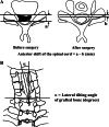
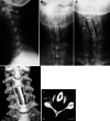
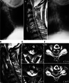
Postoperative C5 palsy is a common complication after cervical spine decompression surgery. However, the incidence, prognosis, and etiology of C5 palsy after anterior decompression with spinal fusion (ASF) have not yet been fully established. In the present study, we analyzed the clinical and radiological characteristics of patients who developed C5 palsy after ASF for cervical degenerative diseases. The cases of 199 consecutive patients who underwent ASF were analyzed to clarify the incidence of postoperative C5 palsy. We also evaluated the onset and prognosis of C5 palsy. The presence of high signal changes (HSCs) in the spinal cord was analyzed using T2-weighted magnetic resonance images. C5 palsy occurred in 17 patients (8.5%), and in 15 of them, the palsy developed after ASF of 3 or more levels. Among ten patients who had a manual muscle test (MMT) grade ≤2 at the onset, five patients showed incomplete or no recovery. Sixteen of the 17 C5 palsy patients presented neck and shoulder pain prior to the onset of muscle weakness. In the ten patients with a MMT grade ≤2 at the onset, nine patients showed HSCs at the C3-C4 and C4-C5 levels. The present findings demonstrate that, in most patients with severe C5 palsy after ASF, pre-existing asymptomatic damage of the anterior horn cells at C3-C4 and C4-C5 levels may participate in the development of motor weakness in combination with the nerve root lesions that occur subsequent to ASF. Thus, when patients with spinal cord lesions at C3-C4 and C4-C5 levels undergo multilevel ASF, we should be alert to the possible occurrence of postoperative C5 palsy.
Figures


Comment in
-
Letter to the Editor concerning "C5 palsy following anterior decompression and spinal fusion for cervical degenerative diseases" by Hashimoto M et al. (2010) Eur Spine J 19:1702-1710.Eur Spine J. 2011 Jul;20(7):1188-9; author reply 1190-1. doi: 10.1007/s00586-010-1674-5. Epub 2011 Jan 4. Eur Spine J. 2011. PMID: 21203891 Free PMC article. No abstract available.
Similar articles
-
Clinical and radiographic analysis of c5 palsy after anterior cervical decompression and fusion for cervical degenerative disease.J Spinal Disord Tech. 2014 Dec;27(8):436-41. doi: 10.1097/BSD.0b013e31826a10b0. J Spinal Disord Tech. 2014. PMID: 22832559
-
Letter to the Editor concerning "C5 palsy following anterior decompression and spinal fusion for cervical degenerative diseases" by Hashimoto M et al. (2010) Eur Spine J 19:1702-1710.Eur Spine J. 2011 Jul;20(7):1188-9; author reply 1190-1. doi: 10.1007/s00586-010-1674-5. Epub 2011 Jan 4. Eur Spine J. 2011. PMID: 21203891 Free PMC article. No abstract available.
-
Spinal cord float back is not an independent predictor of postoperative C5 palsy in patients undergoing posterior cervical decompression.Spine J. 2020 Feb;20(2):266-275. doi: 10.1016/j.spinee.2019.09.017. Epub 2019 Sep 19. Spine J. 2020. PMID: 31542474
-
The incidence of C5 palsy after multilevel cervical decompression procedures: a review of 750 consecutive cases.Spine (Phila Pa 1976). 2012 Feb 1;37(3):174-8. doi: 10.1097/BRS.0b013e318219cfe9. Spine (Phila Pa 1976). 2012. PMID: 22293780 Review.
-
C5 nerve root palsy following decompression of cervical spine with anterior versus posterior types of procedures in patients with cervical myelopathy.Eur Spine J. 2016 Jul;25(7):2050-9. doi: 10.1007/s00586-016-4567-4. Epub 2016 Apr 19. Eur Spine J. 2016. PMID: 27095700 Review.
Cited by
-
Clinical analysis of C5 palsy after cervical decompression surgery: relationship between recovery duration and clinical and radiological factors.Eur Spine J. 2017 Apr;26(4):1101-1110. doi: 10.1007/s00586-016-4664-4. Epub 2016 Jun 24. Eur Spine J. 2017. PMID: 27342613
-
C5 Nerve Palsy After Posterior Instrumentation and Decompression in Cervical Spine Surgery: A Review of the Literature.Cureus. 2025 May 19;17(5):e84430. doi: 10.7759/cureus.84430. eCollection 2025 May. Cureus. 2025. PMID: 40539182 Free PMC article. Review.
-
A Comparative Study on Functional Recovery, Complications, and Changes in Inflammatory Factors in Patients with Thoracolumbar Spinal Fracture Complicated with Nerve Injury Treated by Anterior and Posterior Decompression.Med Sci Monit. 2019 Feb 12;25:1164-1168. doi: 10.12659/MSM.912332. Med Sci Monit. 2019. PMID: 30753178 Free PMC article.
-
Computed tomographic evaluation of C5 root exit foramen in patients with cervical spondylotic myelopathy.Surg Neurol Int. 2014 Apr 16;5(Suppl 3):S59-61. doi: 10.4103/2152-7806.130668. eCollection 2014. Surg Neurol Int. 2014. PMID: 24843812 Free PMC article.
-
Radiographic Measures of Spinal Alignment Are Not Predictive of the Development of C5 Palsy Following Anterior Cervical Discectomy and Fusion Surgery.Int J Spine Surg. 2021 Apr;15(2):213-218. doi: 10.14444/8029. Epub 2021 Apr 1. Int J Spine Surg. 2021. PMID: 33900977 Free PMC article.
References
Publication types
MeSH terms
LinkOut - more resources
Full Text Sources
Medical
Miscellaneous

