

Ultrasound of the knee during voluntary quadriceps contraction: a technique for detecting otherwise occult effusions
- PMID: 20461790
- PMCID: PMC5596890
- DOI: 10.1002/acr.20047
Ultrasound of the knee during voluntary quadriceps contraction: a technique for detecting otherwise occult effusions
Abstract
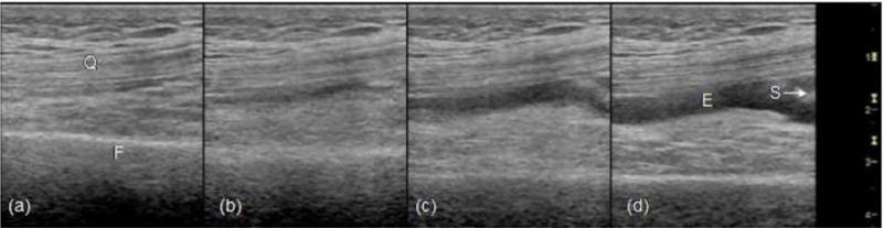
Objective: To describe 1) a technique that can detect synovial effusions not seen on static ultrasound (US) examination and 2) the characteristics of patients with knee osteoarthritis (OA) for whom this technique proved useful.
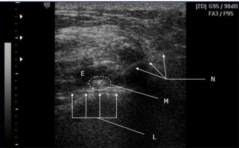
Methods: From reviewed records of 76 patients with knee OA (112 knees) that we had seen for US-guided injections over a defined period, we found 45 knees with no detectable effusion on static US, of which 18 (14 patients) showed fluid when scanned during voluntary quadriceps contraction. For all patients, we had recorded effusion features (physical examination, presence and size on US), and success of joint entry was determined by getting synovial fluid and/or seeing an air echo or inflow of injected material.
Results: The 14 patients we studied were obese (mean +/- SEM body mass index 32.7 +/- 2.3 kg/m(2); 3 morbidly obese), with moderate to severe OA by radiography in most (Kellgren/Lawrence class 3 or 4 in 10 of 14 knees for which radiographs were available). The suprapatellar synovial space seen by US was small (mean +/- SEM depth 0.38 +/- 0.04 cm). Arthrocentesis obtained 0.5-16 ml of synovial fluid (mean +/- SEM 2.9 +/- 0.6 ml), which correlated with the depth of effusion as seen on US with the quadriceps in maximum contraction (Spearman's rho = 0.5597, P = 0.0157). In 4 knees where arthrocentesis failed to retrieve fluid, we observed at injection the inflow of material and a linear air echo.
Conclusion: US of the knee during voluntary quadriceps contraction can find effusions not detectable on static US. Such effusions provide targets for accurate aspiration and injection that would not be appreciated with static US.
Figures
Similar articles
-
Identification of Knee Effusions With Ultrasound: A Comparison of Three Methods.Clin J Sport Med. 2022 Jan 1;32(1):e19-e22. doi: 10.1097/JSM.0000000000000823. Clin J Sport Med. 2022. PMID: 32032167
-
Which knee and probe position determines the final diagnosis of knee inflammation by ultrasound? Results from a European multicenter study.Ultraschall Med. 2012 Dec;33(7):E173-E178. doi: 10.1055/s-0031-1281973. Epub 2011 Dec 22. Ultraschall Med. 2012. PMID: 22194046
-
Accuracy of blind versus ultrasound-guided suprapatellar bursal injection.J Clin Ultrasound. 2012 Jan;40(1):20-5. doi: 10.1002/jcu.20890. Epub 2011 Oct 28. J Clin Ultrasound. 2012. PMID: 22033897 Clinical Trial.
-
Does ultrasound guidance improve the outcomes of arthrocentesis and corticosteroid injection of the knee?Scand J Rheumatol. 2012 Feb;41(1):66-72. doi: 10.3109/03009742.2011.599071. Epub 2011 Nov 21. Scand J Rheumatol. 2012. PMID: 22103390 Clinical Trial.
-
Effect of ultrasound-detected synovitis on therapeutic efficacy of hyaluronic acid injection for symptomatic knee osteoarthritis.Rheumatology (Oxford). 2021 Oct 2;60(10):4486-4494. doi: 10.1093/rheumatology/keab020. Rheumatology (Oxford). 2021. PMID: 33493323 Free PMC article.
Cited by
-
The influence of long distance running on sonographic joint and tendon pathology: results from a prospective study with marathon runners.BMC Musculoskelet Disord. 2016 Jul 11;17:272. doi: 10.1186/s12891-016-1121-9. BMC Musculoskelet Disord. 2016. PMID: 27400865 Free PMC article.
-
Improvement in diagnostic and therapeutic arthrocentesis via constant compression.Clin Rheumatol. 2018 Aug;37(8):2251-2259. doi: 10.1007/s10067-017-3836-x. Epub 2017 Sep 14. Clin Rheumatol. 2018. PMID: 28913649
-
Ultrasonographic Morphological Changes in the Prefemoral Fat Pad Associated with Knee Osteoarthritis.J Med Ultrasound. 2018 Apr-Jun;26(2):94-99. doi: 10.4103/JMU.JMU_15_17. Epub 2018 Jun 12. J Med Ultrasound. 2018. PMID: 30065527 Free PMC article.
-
Differential Diagnosis of Inflammatory Arthropathies by Musculoskeletal Ultrasonography: A Systematic Literature Review.Front Med (Lausanne). 2020 May 7;7:141. doi: 10.3389/fmed.2020.00141. eCollection 2020. Front Med (Lausanne). 2020. PMID: 32457913 Free PMC article.
-
Functional status of the articularis genus muscle in individuals with knee osteoarthritis.J Musculoskelet Neuronal Interact. 2016 Dec 14;16(4):348-354. J Musculoskelet Neuronal Interact. 2016. PMID: 27973387 Free PMC article.
References
-
- Balint PV, Kane D, Hunter J, McInnes IB, Field M, Sturrock RD. Ultrasound guided versus conventional joint and soft tissue aspiration in rheumatology practice: a pilot study. J Rheumatol. 2002;29:2209–13. - PubMed
-
- Kane D, Balint PV, Sturrock RD. Ultrasonography is superior to clinical examination in the detection and localization of knee joint effusion in rheumatoid arthritis. J Rheumatol. 2003;30:966–71. - PubMed
-
- Luck LJ. Musculoskeletal ultrasound intervention: principles and advances. Radiol Clin N Am. 2008;46:515–33. - PubMed
-
- Qvistgaard E, Kristoffersen H, Terslev L, Danneskiold-Samsoe B, Torp-Pedersen S, Bliddal H. Guidance by ultrasound of intra-articular injections in the knee and hip joints. Osteoarthritis Cartilage. 2001;9:512–17. - PubMed
-
- Koski JM, Hermunen HS, Kilponen VM, Saarakkala SJ, Hakulinen UK, Heikkinen JO. Verification of palpation-guided intra-articular injections using glucocorticoid-air-saline mixture and ultrasound imaging (GAS-graphy) Clin Exp Rheumatol. 2006;24:247–52. - PubMed
Publication types
MeSH terms
Grants and funding
LinkOut - more resources
Full Text Sources
