. 2010 Jun 29;49(25):5244-57.
doi: 10.1021/bi100391w.

Extensive and varied modifications in histone H2B of wild-type and histone deacetylase 1 mutant Neurospora crassa

Affiliations

Extensive and varied modifications in histone H2B of wild-type and histone deacetylase 1 mutant Neurospora crassa

D C Anderson et al. Biochemistry. .

Abstract

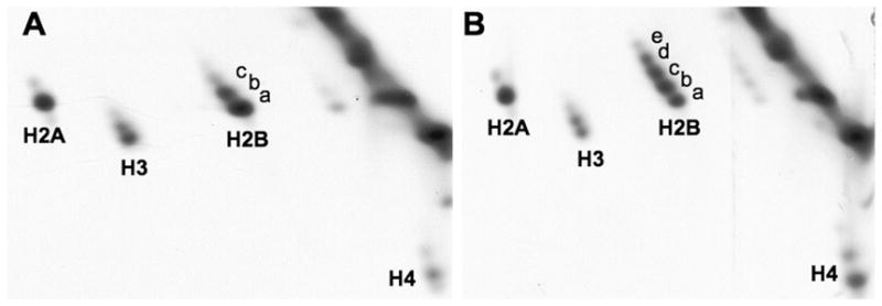
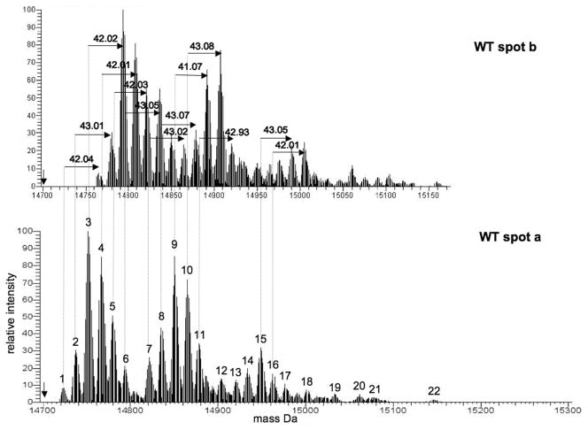
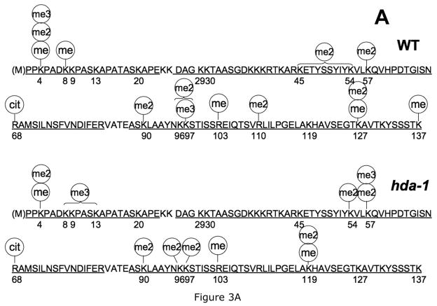
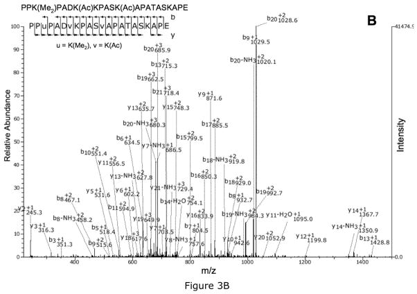
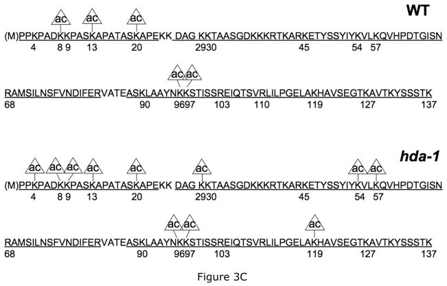
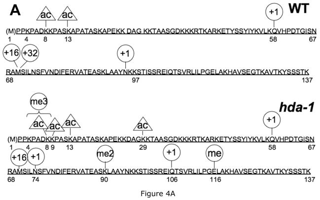
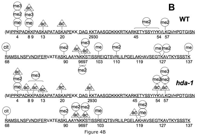
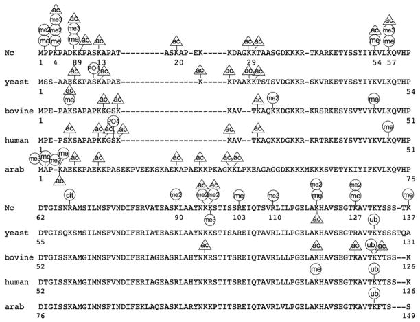
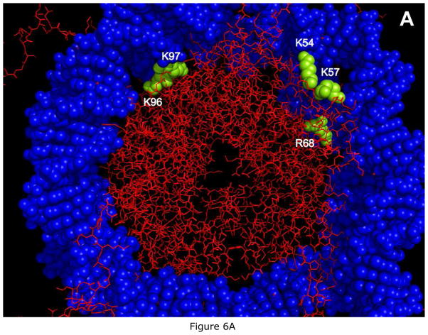
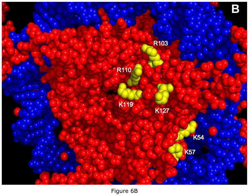
DNA methylation is deficient in a histone deacetylase 1 (HDA1) mutant (hda-1) strain of Neurospora crassa with inactivated histone deacetylase 1. Difference two-dimensional (2D) gels identified the primary histone deacetylase 1 target as histone H2B. Acetylation was identified by LC-MS/MS at five different lysines in wild-type H2B and at 11 lysines in hda-1 H2B, suggesting Neurospora H2B is a complex combination of different acetylated species. Individual 2D gel spots were shifted by single lysine acetylations. FTICR MS-observed methylation ladders identify an ensemble of 20-25 or more modified forms for each 2D gel spot. Twelve different lysines or arginines were methylated in H2B from the wild type or hda-1; only two were in the N-terminal tail. Arginines were modified by monomethylation, dimethylation, or deimination. H2B from wild-type and hda-1 ensembles may thus differ by acetylation at multiple sites, and by additional modifications. Combined with asymmetry-generated diversity in H2B structural states in nucleosome core particles, the extensive modifications identified here can create substantial histone-generated structural diversity in nucleosome core particles.


Figures

Figure 1
Figure 1. High resolution two-dimensional polyacrylamide gel electrophoresis of histones isolated from wild type and hda-1 strains of Neurospora crassa
Histones from wild type (A) and hda-1 (B) strains of Neurospora crassa were resolved in acetic acid-urea-Triton X-100 (AUT) in the horizontal dimension and acetic acid-urea (AU) in the vertical dimension, with the direction of protein migration indicated by the arrows. Proteins were stained with Coomassie blue R-250. All four core histones (H2A, H2B, H3, H4) were observed. Wild type H2B appears as a diagonal ladder of 3 labeled spots with a fourth faint spot, while hda-1 H2B appears as a diagonal ladder of five spots.
Figure 2
Figure 2. FTICR mass spectrometry of electroeluted histone H2B 2D gel spots
The monoisotopic mass of unmodified H2B is 14700.97 Da (vertical arrow). Each single spectral peak represents an isotope cluster for one or more modified forms of histone H2B. Listed mass shifts are based on monoisotopic masses calculated by THRASH (34), which are aligned with the most abundant isotopic peak of each isotope cluster for clarity. The spectra are aligned to allow comparison of clusters of peaks. The high resolution mass spectrum is shown for the lowest (WT spot a) and next upper (WT spot b) wild type histone H2B spots. Mass shifts for each peak are in Table 1. The y-axis represents relative abundance.
Figure 3
Figure 3. Post-translational modifications identified on Neurospora histone H2B
A. Lysine and arginine modifications identified in Neurospora histone H2B. Modifications are indicated by circles above the modified H2B residue (me, methyl; me2, dimethyl; me3, trimethyl; ac, acetyl, cit, arg modified to citrulline). The top sequence indicates modification sites in wild type (WT) H2B; the lower sequence shows modification sites in hda-1 H2B. In some cases MS/MS spectra could not distinguish which of several alternative lysines are methylated; several of these sites are indicated with brackets spanning the potential modified lysines. The sequence coverage in combined LC/MS/MS experiments is indicated by underlined residues. B. MS/MS spectrum identifying the doubly acetylated, doubly methylated hda-1 H2B N-terminal peptide PPK(Me2)PADK(Ac)KPASK(Ac)APATASKAPE using SEQUEST C. Summary of lysine acetylation in wild type and hda-1 H2B. Acetylation at each of the sites is partial since other modifications (or no modification) were identified at each of the indicated sites. The sequence coverage from individual peptides is underlined. Acetylated sites in hda-1 H2B but not in WT H2B are potential substrate sites for the HDA1 histone deacetylase.
Figure 4
Figure 4. Comparison of Neurospora histone H2B post-translational modifications identified by two algorithms
A. Modifications identified by the blind search algorithm InsPecT (28). InsPecT identified several modifications not observed in standard SEQUEST searches, including deamidated asn or gln (circle with +1), met sulfone (circle with +32), and a glutamate methyl ester (me). Other modifications were oxidized met (circle with +16), dimethyl lys (me2), trimethyl lys (me3), and acetyl-lys (ac). Sequence coverage of identified peptides is indicated by underlined residues. Acetyl- and trimethyllysine were distinguished by precursor ion mass. B. Summary of all modifications identified in wild type and HDA1-inactivated H2B using SEQUEST, indicated by circles over modified residues: methyl (me), dimethyl (me2), trimethyl (me3), acetyl (ac), and arg modified to citrulline (cit).
Figure 5
Figure 5. Comparison of Neurospora histone H2B post-translational modifications with those of histone H2B from other organisms
H2B sequences from Neurospora, yeast (Saccharomyces cerevisiae), calf (Bos taurus), Arabidopsis variant HTB11, and human histone H2B (locus CAB02545) were aligned using T-coffee (59). Numbering starts with the N-terminal met although this is not present in the mature histone. Modifications from wild type and HDA1-inactivated H2B were combined for the Neurospora map and include 3 additional modifications reported by Xiong et al (54). The arrow under the Neurospora H2B sequence indicates the start of the globular domain.
Figure 6
Figure 6. Predicted location of post-translationally modified Neurospora histone H2B residues in the crystal structure of the Xenopus nucleosome core particle
Positions of the post-translationally modified Neurospora H2B globular domain residues in the structure of the Xenopus nucleosome core particle (PDB1KX5) were derived by sequence alignment (59) with Xenopus H2B (Table 8). Residues on only one face of the nucleosome core particle structure are shown; similar residues are also on the opposite face. Histone polypeptides are red; modified residues are yellow, and the two strands of DNA wrapped around the histone octamer are blue. A. H2B modified residues predicted to be close to DNA and which may directly or indirectly bind the DNA wrapped around the core histone octamer. K96, K97 and R68 are buried in the histone octamer structure while K54 and K57 are both at the surface of the NCP. B. H2B residues predicted to be on or near the surface of the nucleosome core particle (NCP). Numbers are shown for Neurospora H2B residues.

Similar articles

Cited by

References

    1. McGhee JD, Felsenfeld G. Nucleosome structure. Ann Rev Biochem. 1980;49:1115–1156. - PubMed
    1. Strahl B, Allis C. The language of covalent histone modifications. Nature. 2000;403:41–45. - PubMed
    1. Green GR. Phosphorylation of histone variant regions in chromatin: Unlocking the linker? Biochemistry and Cell Biology. 2001;79:275–287. - PubMed
    1. Fischle W, Wang Y, Allis CD. Binary switches and modification cassettes in histone biology and beyond. Nature. 2003;425:475–479. - PubMed
    1. Zhang L, Eugeni E, Parthun MR, Freitas M. Identification of novel histone post-translational modifications by peptide mass fingerprinting. Chromosoma. 2003;112:77–86. - PubMed

Publication types

MeSH terms