. 2010;12(3):R27.
doi: 10.1186/bcr2575. Epub 2010 May 12.

Inhibition of phosphatidylcholine-specific phospholipase C downregulates HER2 overexpression on plasma membrane of breast cancer cells

Affiliations

Inhibition of phosphatidylcholine-specific phospholipase C downregulates HER2 overexpression on plasma membrane of breast cancer cells

Luisa Paris et al. Breast Cancer Res. 2010.

Abstract

Introduction: Overexpression on plasma membrane of human epidermal growth factor receptor 2 (HER2) is reported in 25% to 30% of breast cancers. Heterodimer formation with cognate members of the epidermal growth factor receptor (EGFR) family, such as HER3 and EGFR, activates abnormal cell-signalling cascades responsible for tumorigenesis and further transcriptional HER2 gene upregulation. Targeting the molecular mechanisms controlling HER2 overexpression and recycling may effectively deactivate this feedback-amplification loop. We recently showed that inactivation of phosphatidylcholine-specific phospholipase C (PC-PLC) may exert a pivotal role in selectively modulating the expression on the membrane of specific receptors or proteins relevant to cell function. In the present study, we investigated the capability of PC-PLC inhibition to target the molecular mechanisms controlling HER2 overexpression on the membrane of breast cancer cells by altering the rates of its endocytosis and lysosomal degradation.

Methods: Localization on the membrane and interaction of PC-PLC with HER2, EGFR, and HER3 were investigated on HER2-overexpressing and HER2-low breast cancer cell lines, by using confocal laser scanning microscopy, flow cytometry, cell-surface biotinylation, isolation of lipid rafts, and immunoprecipitation experiments. The effects of the PC-PLC inhibitor tricyclodecan-9-yl-potassium xanthate (D609) on HER2 expression on the membrane and on the levels of overall HER2, HER2-HER3, and HER2-EGFR contents were monitored in the HER2-overexpressing SKBr3 cells, after either transient or continuous receptor engagement with anti-HER2 monoclonal antibodies, including trastuzumab. Changes of HER2 expression and cell proliferation were examined in SKBr3, BT-474, and MDA-MB-453 cells continuously exposed to D609 alone or combined with trastuzumab.

Results: PC-PLC selectively accumulates on the plasma membrane of HER2-overexpressing cells, where it colocalizes and associates with HER2 in raft domains. PC-PLC inhibition resulted in enhanced HER2 internalization and lysosomal degradation, inducing downmodulation of HER2 expression on the membrane. Moreover, PC-PLC inhibition resulted in strong retardation of HER2 reexpression on the membrane and a decrease in the overall cellular contents of HER2, HER2-HER3, and HER2-EGFR heterodimers. The PC-PLC inhibitor also induced antiproliferative effects, especially in trastuzumab-resistant cells.

Conclusions: The results pointed to PC-PLC inhibition as a potential means to counteract the tumorigenic effects of HER2 amplification and complement the effectiveness of current HER2-targeting therapies.


Figures

Figure 1
Figure 1
PC-PLC expression on plasma membrane of breast cancer cells. (a) CLSM analyses of six unfixed cell lines stained for PC-PLC detection (pseudo-color grey). Upper panels show examples of a three-dimensional reconstruction of a single cell (scale bars, 5 μm); the lower panels show single central sections of a group of cells (scale bars, 20 μm). Nuclei were stained with DAPI (blue). (b, c) FACS analyses of unfixed cells: representative cytofluorimetric histograms of PC-PLC relative fluorescence intensity of the indicated cell lines (b) and percentage (mean ± SD) of PC-PLC positive cells (c). Statistical significance of differences between HER2-overexpressing (SKBr3 and MDA-MB-453) and HER2-non-overexpressing (MDA-MB-231, MCF-7, MDA-MB-435) carcinoma cell lines was P < 0.001. Five independent experiments were performed on each cell line.
Figure 2
Figure 2
Colocalization of PC-PLC and HER2 on plasma membrane of SKBr3 cells. (a) CLSM detection of PC-PLC and HER2 in either unfixed (top and middle panels) or fixed and permeabilized SKBr3 cells (bottom panel) by using rabbit polyclonal α-PC-PLC (green) and α-HER2 W6/100 mAb (red). Colocalization areas are represented in yellow. The middle panel shows the tridimensional reconstruction of PC-PLC and HER2 expression on the plasma membrane. Scale bars, 8 μm. At least five independent series of experiments were performed for each condition. (b) PC-PLC immunoblotting analyses of immunoprecipitated biotinylated proteins isolated from SKBr3 cells, detected by streptavidin-HRP (left, IB:STREP HRP) or by α-PC-PLC (right, IB:α-PC-PLC). TL, total lysate; bTL, biotinylated total lysate; CTR IgG, control for α-PC-PLC IgG; IP α-PC-PLC, α-PC-PLC immunoprecipitates. *IgG heavy chains. (c) Sucrose gradient fractions isolated from SKBr3 cell lysates and analyzed by Western blotting for HER2 (185 kDa) and PC-PLC (66 kDa) detection. T, total cell lysate. (d, e) Western blot analyses of α-HER2 and α-PC-PLC (d) and α-EGFR and α-PC-PLC (e) immunoprecipitates (IP), blotted with the mutual Abs (α-PC-PLC, α-HER2, and α-EGFR, respectively), compared with the respective controls (CTR IgG α-PC-PLC, CTR IgG α-HER2, CTR IgG α-EGFR). Panels b, c, d, show representative results of three independent experiments. The immunoprecipitation in panel e was repeated twice.
Figure 3
Figure 3
D609-induced PC-PLC inhibition and HER2 downmodulation in SKBr3 cells. (a) Amplex Red® assay SKBr3 cells after exposure to the PC-PLC inhibitor D609. Relative PC-PLC activity was measured in the presence of D609 (50 μg/mL, black columns) and compared with that of untreated control cells (t = 0 taken as 1.0 or t = 72 hours, white columns). Histograms represent the mean ± SD (n = 4). (b) CLSM analyses of unfixed SKBr3 cells cultured in complete medium (t = 0) and in presence of D609 for the indicated time periods. After washing, cells were stained with α-PC-PLC (green) and α-HER2 (red). Nuclei were stained with DAPI (blue). Colocalization areas are represented in yellow. Scale bars, 5 μm. These experiments were independently repeated 3 times.
Figure 4
Figure 4
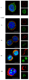
Effect of PC-PLC inhibition on HER2 internalization in SKBr3 cells. CLSM analyses of SKBr3 cells cultured in complete medium either in the absence (t = 0) or presence of D609 (50 μg/mL) for the indicated incubation times. After washing, cells were fixed and stained with α-PC-PLC (green) and α-HER2 Abs (red). Nuclei were stained with DAPI (blue). Colocalization areas were represented in yellow (merge between green and red) or cyan (merge between green and DAPI). Scale bars, 8 μm. Panels show representative examples of five independent series of experiments.
Figure 5
Figure 5
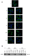
Effects of D609 on trafficking and degradation of HER2 receptor molecules in SKBr3 cells. (a) CLSM analyses of SKBr3 cells, incubated for the indicated time intervals with the PC-PLC inhibitor D609 (50 μg/mL) and then fixed and stained with the α-HER2 mAb W6/100 (red) and α-Rab5B Ab (green, panels a through d) or α-Lamp 2 Ab (green, panels e through h). Nuclei were stained with DAPI (blue). Scale bars, 8 μm. (b) Western blot analyses of total lysates of SKBr3 cells either untreated or incubated with D609 for 6 and 24 hours. The blots were incubated with α-HER2 or α-PC-PLC Abs. Actin was used as quantitative loading control. Experiments shown in panels a and b were independently repeated 3 and 4 times, respectively.
Figure 6
Figure 6
D609-induced retardation of reexpression of HER2 on the plasma membrane of SKBr3 cells after short-term receptor engagement with an anti-HER2 antibody. (a) CLSM examinations on unfixed cells transiently engaged (30 minutes at 4°C) with α-HER2 mAb 300G9, followed by goat α-mouse Alexa Fluor-594 (panel a, red), then cultured at 37°C for the indicated time periods in complete Ab-free medium, either in the absence (b through g) or presence of D609, 50 μg/mL (h through o). At the end of each time interval, cells were stained again on the plasma membrane with the α-HER2 W6/100 mAb, followed by goat α-mouse Alexa Fluor-488 (green). Nuclei were stained with DAPI (blue). Scale bars, 8 μm. These analyses were independently performed 3 times. (b) Western blot analyses of SKBr3 cells first engaged with α-HER2 300G9 mAb, followed by goat α-mouse antibody, and then cultured at 37°C either in the absence or presence of D609 for the indicated time periods. The blots were incubated with α-HER2 or with anti-β actin Abs. These experiments were repeated on seven sets of independent cell preparations.
Figure 7
Figure 7
Effect of D609 on HER2 expression in SKBr3 cells continuously exposed to trastuzumab. (a) CLSM analyses of cells fixed after exposure for the indicated time intervals to trastuzumab, in the presence or absence of D609. Nuclei were stained with DAPI (blue). Scale bars, 20 μm. (b) Decrease in the overall HER2 content, detected by Western blotting, in total lysates of cells after incubation for different time intervals with D609 (50 μg/mL), trastuzumab (HERC, 10 μg/mL), or their combination (COMB). CTR, untreated control cells. Both CLSM and immunoblotting analyses were repeated 3 times.
Figure 8
Figure 8
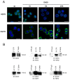
Effects of PC-PLC inhibition on EGFR and HER3 internalization, overall receptors' contents and heterodimer formation with HER2. (a) CLSM analyses of fixed SKBr3 cells after exposure to D609 for different time intervals and stained with either α-HER3 (upper panels) or α-EGFR antibodies (lower panels). Nuclei were stained with DAPI (blue). Scale bars, 20 μm. (b) Immunoblotting (IB) and immunoprecipitation (IP) experiments on SKBr3 cells, incubated with D609 for 24 hours, to detect overall HER3 and EGFR contents (left panels) and their levels of heterodimerization with HER2 (right). Central panels show the efficiency of the α-HER3 and α-EGFR antibodies in immunoprecipitating the respective target receptors. ET CTR, total extract of control cells; ET D609, total extract of D609-treated cells; CTR IgG, control IgG of α-HER3 or α-EGFR antibodies; IP CTR, immunoprecipitation from control cells; IP D609, immunoprecipitation from D609-treated cells. Representative examples of two independent experiments are shown.
Figure 9
Figure 9
Antiproliferative effect of PC-PLC inhibition on HER2-overexpressing cells. MTT assays on (a) SKBr3 and (b) BT-474 cells both exposed to trastuzumab (HERC, 10 μg/mL), or (c) MDA-MB-453 exposed to trastuzumab (50 μg/mL), after incubation for the indicated times with D609, trastuzumab (HERC), or their combination (COMB). The percentage of cell proliferation was calculated by defining the absorption of untreated cells as 100%. Histograms represent the mean percentage of independent series of assays (± SD for SKBr3 and BT-474 (n = 3); or ± maximum deviation for MDA-MB-453 (n = 2)). Significance of differences: **P ≤ 0.02; ***P < 0.005.

Similar articles

Cited by

References

    1. Yarden Y, Sliwkowski MX. Untangling the ErbB signalling network. Nat Rev Mol Cell Biol. 2001;2:127–137. doi: 10.1038/35052073. - DOI - PubMed
    1. Mendelsohn J, Baselga J. Status of epidermal growth factor receptor antagonists in the biology and treatment of cancer. J Clin Oncol. 2003;21:2787–2799. doi: 10.1200/JCO.2003.01.504. - DOI - PubMed
    1. Lemmon MA. The EGF receptor family as therapeutic targets in breast cancer. Breast Dis. 2003;18:33–43. - PubMed
    1. Hynes NE, Lane HA. ERBB receptors and cancer: the complexity of targeted inhibitors. Nat Rev Cancer. 2005;5:341–354. doi: 10.1038/nrc1609. - DOI - PubMed
    1. Carlsson J, Nordgren H, Sjöström J, Wester K, Villman K, Bengtsson NO, Ostenstad B, Lundqvist H, Blomqvist C. HER2 expression in breast cancer primary tumours and corresponding metastases: original data and literature review. Br J Cancer. 2004;90:2344–2348. - PMC - PubMed

Publication types