Enhancement of cisplatin sensitivity in Lewis Lung carcinoma by liposome-mediated delivery of a survivin mutant
- PMID: 20462440
- PMCID: PMC2878293
- DOI: 10.1186/1756-9966-29-46
Enhancement of cisplatin sensitivity in Lewis Lung carcinoma by liposome-mediated delivery of a survivin mutant
Abstract
Background: A high concentration of cisplatin (CDDP) induces apoptosis in many tumor cell lines. CDDP has been administered by infusion to avoid severe toxicity. Recently, it has been reported that changes in survivin expression or function may lead to tumor sensitization to chemical and physical agents. The aim of this study was to determine whether a dominant-negative mouse survivin mutant could enhance the anti-tumor activity of CDDP.
Methods: A plasmid encoding the phosphorylation-defective dominant-negative mouse survivin threonine 34-->alanine mutant (survivin T34A) complexed to a DOTAP-chol liposome (Lip-mS) was administered with or without CDDP in Lewis Lung Carcinoma (LLC) cells and in mice bearing LLC tumors, and the effects on apoptosis, tumor growth and angiogenesis were assessed. Data were analyzed using one-way analysis of variance(ANOVA), and a value of P < 0.05 was considered to be statistically significant.
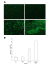
Results: LLC cells treated with a combination of Lip-mS and CDDP displayed increased apoptosis compared with those treated with Lip-mS or CDDP alone. In mice bearing LLC tumors and treated with intravenous injections of Lip-mS and/or CDDP, combination treatment significantly reduced the mean tumor volume compared with either treatment alone. Moreover, the antitumor effect of Lip-mS combined with CDDP was greater than their anticipated additive effects.
Conclusion: These data suggest that the dominant-negative survivin mutant, survivin T34A, sensitized LLC cells to chemotherapy of CDDP. The synergistic antitumor activity of the combination treatment may in part result from an increase in the apoptosis of tumor cells, inhibition of tumor angiogenesis and induction of a tumor-protective immune response.
Figures


Similar articles
-
Enhanced tumor radiosensitivity by a survivin dominant-negative mutant.Oncol Rep. 2010 Jan;23(1):97-103. Oncol Rep. 2010. PMID: 19956869
-
Efficient inhibition of murine breast cancer growth and metastasis by gene transferred mouse survivin Thr34-->Ala mutant.J Exp Clin Cancer Res. 2008 Sep 25;27(1):46. doi: 10.1186/1756-9966-27-46. J Exp Clin Cancer Res. 2008. PMID: 18816410 Free PMC article.
-
Enhancement of cisplatin sensitivity in lung cancer xenografts by liposome-mediated delivery of the plasmid expressing small hairpin RNA targeting Survivin.J Biomed Nanotechnol. 2012 Aug;8(4):633-41. doi: 10.1166/jbn.2012.1419. J Biomed Nanotechnol. 2012. PMID: 22852473
-
Co-delivery of doxorubicin and plasmid by a novel FGFR-mediated cationic liposome.Int J Pharm. 2010 Jun 30;393(1-2):119-26. doi: 10.1016/j.ijpharm.2010.04.018. Epub 2010 Apr 21. Int J Pharm. 2010. PMID: 20416367
-
Inhibition of lymphatic metastases by a survivin dominant-negative mutant.Oncol Res. 2012;20(12):579-87. doi: 10.3727/096504013X13775486749416. Oncol Res. 2012. PMID: 24139416
Cited by
-
Curcumin reduces the expression of survivin, leading to enhancement of arsenic trioxide-induced apoptosis in myelodysplastic syndrome and leukemia stem-like cells.Oncol Rep. 2016 Sep;36(3):1233-42. doi: 10.3892/or.2016.4944. Epub 2016 Jul 15. Oncol Rep. 2016. PMID: 27430728 Free PMC article.
-
Ligand-targeted theranostic nanomedicines against cancer.J Control Release. 2016 Oct 28;240:267-286. doi: 10.1016/j.jconrel.2016.01.002. Epub 2016 Jan 6. J Control Release. 2016. PMID: 26772878 Free PMC article. Review.
-
p62/SQSTM1 levels predicts radiotherapy resistance in hypopharyngeal carcinomas.Am J Cancer Res. 2017 Apr 1;7(4):881-891. eCollection 2017. Am J Cancer Res. 2017. PMID: 28469960 Free PMC article.
-
Nucleic acid cancer vaccines targeting tumor related angiogenesis. Could mRNA vaccines constitute a game changer?Front Immunol. 2024 Jul 16;15:1433185. doi: 10.3389/fimmu.2024.1433185. eCollection 2024. Front Immunol. 2024. PMID: 39081320 Free PMC article. Review.
-
Survivin - biology and potential as a therapeutic target in oncology.Onco Targets Ther. 2013 Oct 16;6:1453-62. doi: 10.2147/OTT.S33374. Onco Targets Ther. 2013. PMID: 24204160 Free PMC article. Review.
References
Publication types
MeSH terms
Substances
LinkOut - more resources
Full Text Sources

