. 2010 Sep;38(17):5944-57.
doi: 10.1093/nar/gkq334. Epub 2010 May 12.

Physical basis of the inducer-dependent cooperativity of the Central glycolytic genes Repressor/DNA complex

Affiliations

Physical basis of the inducer-dependent cooperativity of the Central glycolytic genes Repressor/DNA complex

Denis Chaix et al. Nucleic Acids Res. 2010 Sep.

Abstract

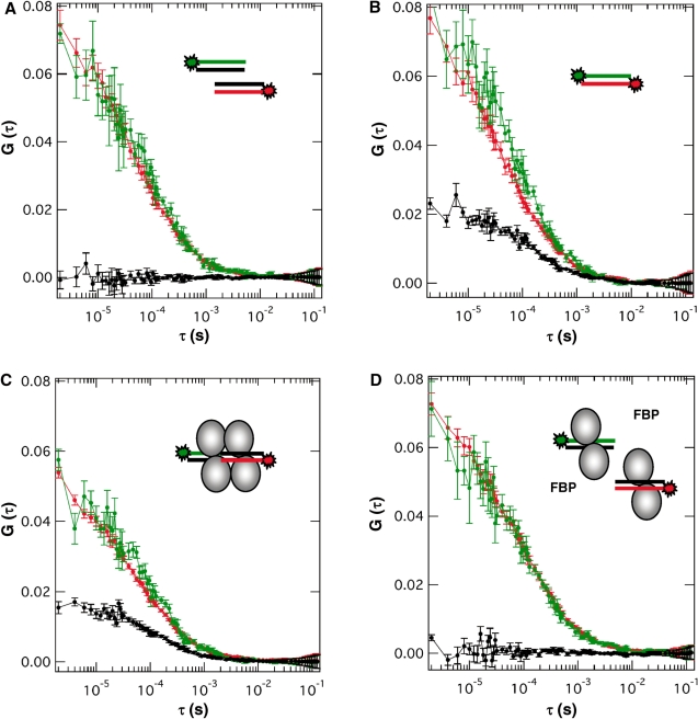
The Central glycolytic genes Repressor (CggR) from Bacillus subtilis belongs to the SorC family of transcription factors that control major carbohydrate metabolic pathways. Recent studies have shown that CggR binds as a tetramer to its tandem operator DNA sequences and that the inducer metabolite, fructose 1,6-bisphosphate (FBP), reduces the binding cooperativity of the CggR/DNA complex. Here, we have determined the effect of FBP on the size, shape and stoichiometry of CggR complexes with full-length and half-site operator sequence by small-angle X-ray scattering, size-exclusion chromatography, fluorescence cross-correlation spectroscopy and noncovalent mass spectrometry (MS). Our results show that CggR forms a compact tetrameric assembly upon binding to either the full-length operator or two half-site DNAs and that FBP triggers a tetramer-dimer transition that leaves a single dimer on the half-site or two physically independent dimers on the full-length target. Although the binding of other phospho-sugars was evidenced by MS, only FBP was found to completely disrupt dimer-dimer contacts. We conclude that inducer-dependent dimer-dimer bridging interactions constitute the physical basis for CggR cooperative binding to DNA and the underlying repression mechanism. This work provides experimental evidences for a cooperativity-based regulation model that should apply to other SorC family members.


Figures

Figure 1.
Figure 1.
The SEC and SAXS data of CggR/DNA complexes. (A) The B. subtilis gapA operator region targeted by the CggR repressor. The nucleotide sequence of the CggR-binding region identified by DNase I footprinting experiment (17) is shown, located 25-bp downstream of the transcription start (+1), and upstream of the translation initiation codon of cggR, the first coding sequence of the hexacistronic gapA operon (15). The full-length operator sequence OLR is composed of two tandem repeats (long arrows), OL and OR, each comprising a small palindrome (small black arrows) proposed to constitute the recognition motif for one CggR dimer (25). Indicated bellow is the length of the DNA fragments and the color code used in Figure 1 (B–D) for representing SEC and SAXS data obtained with the different CggR/DNA complexes in the presence/absence of the inducer metabolite FBP. (B) Elution profiles of 250 µl samples of purified CggR/DNA complexes (colored curves) at 1 mg/ml (protein concentration) and injected on Superdex 200 HR10/30 (GE Healthcare) at a flow rate of 0.5 ml/min in 150 mM NaCl, 20 mM Tris-HCl pH 8, 2 mM EDTA, 2 mM DTT, eventually supplemented with 0.5 mM FBP (red and green curves). Black curves, dsDNA alone corresponding to the full-length operator (OLR, 45 bp, dotted line) or the right half-site (OR, 23 bp, plain line). (C) Comparison of the normalized scattering profiles of CggR/DNA complexes in the presence and absence of FBP [identical samples and buffer condition as in (B)]. (D) Distance distribution function, P(r), computed from the experimental scattering data and normalized to the maximum value of unity using GNOM (39).
Figure 2.
Figure 2.
Auto- and cross-correlation profiles recovered from FCCS measurements with Atto-647N-labeled or fluorescein-labeled oligonucleotides corresponding to the CggR OR half-site operator in the presence/absence of the repressor and the inducer metabolite FBP. Schematics of the labeled DNAs and protein present in the sample chambers are shown. Red, blue and black curves correspond to the autocorrelation traces recorded in the red (675 nm) and blue (525 nm) detection channels and the cross-correlation curve, respectively. (A) A mixture of the singly labeled dsDNA fragments showing the absence of cross-correlation signal. (B) A doubly labeled DNA hybrid serving as positive control of cross-correlation. The difference in the maximum amplitude of the auto- and cross-correlation function (G(0)) denotes the partial labeling and hybridization of the two labeled DNA strands. (C) Cross-correlation upon addition of the repressor protein to the singly labeled DNA mixture of panel (A), evidencing the CggR-mediated assembly of the DNA fragments. (D) Loss of cross correlation signal upon addition of the inducer metabolite to the protein/DNA mixture of panel (C), evidencing the FBP-induced disruption of the CggR/DNA ternary complex. Concentration was 60 nM for the labeled DNA fragments, 300 nM for CggR (monomer unit) and 0.5 mM for FBP.
Figure 3.
Figure 3.
Noncovalent MS analysis of CggR interaction with the OR half-operator DNA and assessment of inducer binding specificity. The CggR/OR complex was diluted to 3 µM in 150 mM ammonium acetate buffer at pH 8.0 and analyzed either (A) alone or in presence of 30 µM (B) FBP, (C) F6P or (D) GBP. Chemical representations of the phospho-sugars are shown on the left. Insets on the right represent enlarged mass spectra showing the +20 charge state of the 2:1 CggR/OR complex and the +23 charge state of the 4:2 CggR/OR complex. Circled numbers correspond to the number of phospho-sugar molecules bound to each complex. Similar results were obtained when adding the phospho-sugars at 60 µM (data not shown).
Figure 4.
Figure 4.
SASREF rigid body models and their fit (magenta line) to experimental SAXS data (dots) of the CggR complexes with the half-site operator DNA OR (A and D) or the full-length operator OLR (B and C) and in the presence (A and B) or absence (C and D) of FBP. The 1Z9C-derived models of the dimeric or tetrameric N-terminal wH domain of CggR (whCggR) bound to the OR or OLR dsDNA are colored in dark blue, with the DNA shown as sticks and the whCggR dimers in surface representation. Red, cyan, yellow and green molecular surfaces represent the rdCggR that moved as independent units during rigid body modeling by SASREF (48). The experimental chi value (χe) of the fitting curves given by SASREF as well as the theoretical gyration radius (Rg) and Stokes’ radius (Rs) of the models calculated by HYDROPRO (49) are indicated in the bottom-left corner of the graphics. Shown here are representative examples of at least 10 different models generated automatically for each complex, combining one of the best fit and agreement with experimentally determined size parameters given in Table 1. Two orientations of the dimeric complex are shown in (A). Panels (C) and (D) purposely exemplify two different models that fit equally well the SAXS data of the tetrameric complex in the absence of FBP.
Figure 5.
Figure 5.
Structural mechanism for CggR-mediated regulation of the gapA operon. In the proposed model, transcriptional regulation by the CggR repressor and its inducer metabolite FBP is achieved by modulating the extent of cooperative interactions rather than simply DNA binding. (A) In the absence of glucose or other carbon sources producing high levels of FBP, CggR interacts as a dimer of dimers bridging the two half-sites of the gapA operator region. This tetrameric assembly can efficiently block the progression of the RNA polymerase, thereby leading to the arrest of transcription. (B) In the presence of glucose, the raise of FBP intracellular concentration provokes the disruption of dimer–dimer contacts through inducer binding to the low-affinity sugar-binding site of the CggR repressor. CggR dimers can still bind independently to each half-operator, however with much lower affinity for the left (5′) half-site than for the right (3′) half-site. This provides a higher opportunity for the transcribing RNA polymerase to read through the first then the second half-site, thereby leading to transcription of the downstream coding sequences.

Similar articles

Cited by

References

    1. Shea MA, Ackers GK. The OR control system of bacteriophage lambda. A physical-chemical model for gene regulation. J. Mol. Biol. 1985;181:211–230. - PubMed
    1. Grillo AO, Brown MP, Royer CA. Probing the physical basis for trp repressor-operator recognition. J. Mol. Biol. 1999;287:539–554. - PubMed
    1. Chahla M, Wooll J, Laue TM, Nguyen N, Senear DF. Role of protein-protein bridging interactions on cooperative assembly of DNA-bound CRP-CytR-CRP complex and regulation of the Escherichia coli CytR regulon. Biochemistry. 2003;42:3812–3825. - PubMed
    1. Beckett D. Linked equilibria in regulation of transcription initiation. Methods Cell Biol. 2008;84:25–52. - PubMed
    1. Connaghan KD, Heneghan AF, Miura MT, Bain DL. Na(+) and K(+) Allosterically regulate cooperative DNA binding by the human progesterone receptor. Biochemistry. 2010;49:422–431. - PMC - PubMed

Publication types