. 2010 Sep 1;87(4):618-27.
doi: 10.1093/cvr/cvq141. Epub 2010 May 11.

Blockade of self-reactive IgM significantly reduces injury in a murine model of acute myocardial infarction

Affiliations

Blockade of self-reactive IgM significantly reduces injury in a murine model of acute myocardial infarction

Michael S Haas et al. Cardiovasc Res. .

Abstract

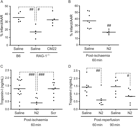
Aims: Coronary artery occlusion resulting in ischaemia/reperfusion (I/R) injury is a major cause of mortality in the western world. Circulating natural IgM has been shown to play a significant role in reperfusion injury, leading to the notion of a pathogenic response against self by the innate immune system. A specific self-antigen (non-muscle myosin heavy chain II) was recently identified as the major target of pathogenic natural IgM. Therefore, we hypothesized that a synthetic peptide mimetope (N2) or monoclonal antibodies directed against the self-antigen would prevent specific IgM binding to the self-antigen and reduce reperfusion injury in the heart.

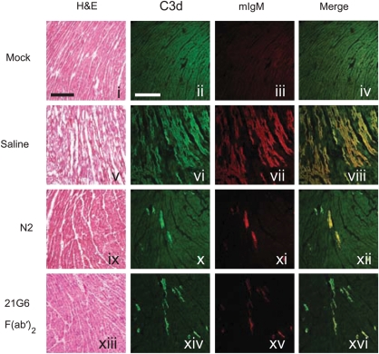
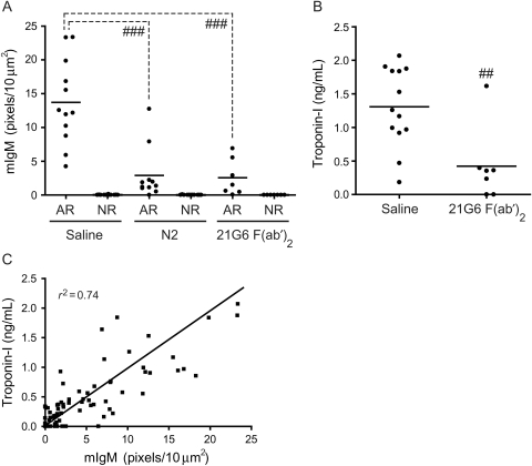
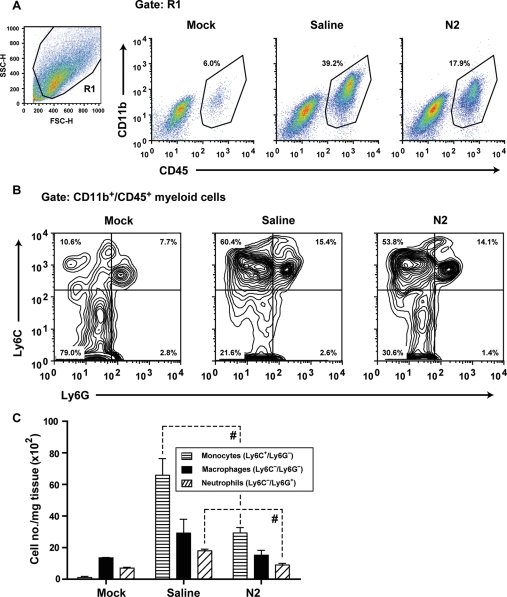
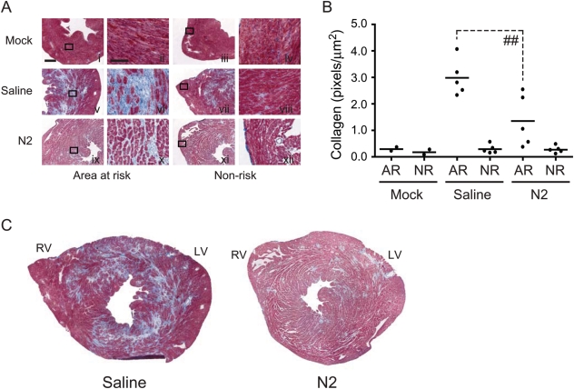
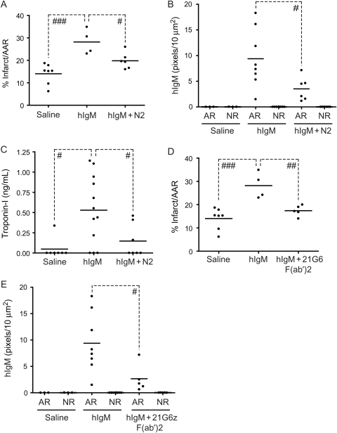
Methods and results: We find that treatment with N2 peptide reduces infarct size by 47% and serum cardiac troponin-I levels by 69% following 1 h ischaemia and 24 h reperfusion. N2 peptide or an anti-N2 F(ab')(2) (21G6) is also effective at preventing IgM and complement deposition. Additionally, N2 peptide treatment significantly reduces monocyte and neutrophil infiltration at 24 h and collagen deposition at 5 days. Finally, we show that human IgM (hIgM) also includes specificity for the highly conserved self-antigen and that myocardial injury in antibody-deficient mice reconstituted with hIgM is blocked by treatment with N2 peptide or 21G6 F(ab')(2).

Conclusion: The findings in this study identify potential therapeutics [i.e. N2 peptide or 21G6 F(ab')(2)] that prevent specific IgM binding to ischaemic antigens in the heart, resulting in a significant reduction in cardiac I/R injury.


Figures

Figure 1
Figure 1
Effect of N2 peptide on myocardial I/R injury. (A) IgM from a single hybridoma clone (IgMcm-22) restores myocardial I/R injury in RAG-1−/− mice. RAG-1−/− mice were injected iv with 200 µg of IgMcm-22 and then subjected to I/R. Infarct sizes were determined by TTC staining. (B and C) Effect of N2 peptide on infarct size (B) and cTnI levels (C). N2 or N2 scrambled (Scr) peptide (200 µg) was injected iv into C57BL/6 mice following 60 min ischaemia (post-ischaemia). Infarct sizes were determined after 24 h reperfusion by TTC staining (B) and serum cTnI levels were also measured (C). (D) Effect of N2 peptide during early stages of reperfusion. C57BL/6 mice were first subjected to 60 min ischaemia after which N2 peptide (200 µg) was injected at various times during reperfusion (post-reperfusion). After 24 h of total reperfusion, serum cTnI levels were analysed (#P < 0.05; ##P < 0.01; ###P < 0.001).
Figure 2
Figure 2
Effect of N2 peptide and 21G6 F(ab′)2 on myocardial I/R injury. Representative cryosections of mock-, NS-, N2-, or 21G6 F(ab′)2-treated C57BL/6 mice subjected to I/R. Cryosections were stained with H&E, anti-C3d-FITC, and anti-IgM-568. Scale bars, 200 µm.
Figure 3
Figure 3
(A) Quantitation of IgM staining in the area of risk (AR) and non-risk (NR) area of NS-, N2-, and 21G6 F(ab′)2-treated C57BL/6 mice. (B) Effect of 21G6 F(ab′)2 on cTnI levels following I/R. 21G6 F(ab′)2 (150 µg) was injected iv into C57BL/6 mice following 60 min ischaemia and serum cTnI levels were measured 24 h post-ischaemia. Each filled circle represents a single mouse (##P < 0.01; ###P < 0.001). Images were collected with a Zeiss/BioRad Radiance 2000MP system attached to an Olympus BX50WI upright microscope, ×20 UPlanapo N.A. 0.7 objective, controlled by the Lasersharp 2000 software. (C) Correlation between serum cTnI levels and IgM deposition in C57BL/6 mice.
Figure 4
Figure 4
Effect of N2 treatment on neutrophil and monocyte infiltration of injured myocardium. C57BL/6 mice were injected with NS or N2 peptide and subjected to I/R. Inflammatory cell populations were analysed by FACS of mock-, NS-, or N2-treated hearts. (A) Representative FACS plots show myeloid cells in the heart, defined by expression of CD45 and CD11b. The live-cell gate is shown on the left (forward vs. side scatter). (B) Gating on myeloid cells (CD11b+/CD45+), three distinct populations were defined: infiltrating monocytes (Ly6C+/Ly6G), resident macrophages (Ly6C/Ly6G), and neutrophils (Ly6C/Ly6G+). Corresponding percentages of each population are indicated on plots. (C) Cumulative analyses of heart inflammation. N2 peptide treatment significantly decreases both infiltrating monocyte and neutrophil populations. Absolute leucocyte numbers were calculated from total cell count, relative population proportions within each heart, and tissue weight (#P < 0.05).
Figure 5
Figure 5
Effect of N2 treatment on collagen deposition in C57BL/6 mice. (A) C57BL/6 mice were injected iv with NS or N2 peptide and subjected to 1 h ischaemia and 5-day reperfusion. Hearts were collected after 5-day reperfusion and cryosections were stained with MT to evaluate collagen content. (B) Quantitation of collagen in the AR and NR area of NS- and N2-treated C57BL/6 mice. Each filled circle represents a single mouse. (C) Representative composite MT-stained cryosections from either NS- or N2-treated C57BL/6 mice. Scale bar shown in panel i, 500 µm; panel ii, 100 µm (##P < 0.01). Images were collected with a Leica DMLB, ×20 AR N.A. 0.5 Leica objective and captured with a Leica DFP480 camera. Photoshop Version 7 software was used for quantitation.
Figure 6
Figure 6
Reconstitution of RAG-1−/− mice with hIgM. (A) RAG-1−/− mice were injected iv with pooled individual hIgM (400 µg) from 10 donors followed by NS or N2 peptide and subjected to I/R. Infarct sizes were determined by TTC staining. (B and C) RAG-1−/− mice were treated as in (A) and analysed for hIgM deposition (B) or serum cTnI levels (C). (D and E) RAG-1−/− mice were injected iv with hIgM as in (A) and then NS or 21G6 F(ab′)2 and subjected to I/R. Infarct sizes were determined by TTC staining (D) or analysed for hIgM deposition (E) (#P < 0.05; ##P < 0.01; ###P < 0.001).

Comment in

Similar articles

Cited by

References

    1. Kumar V, Abbas AK, Fausto N, Aster JC. Robbins and Contran Pathologic Basis of Disease. Professional Edition. 8th ed. Philadelphia, PA: Saunders Elsevier; 2010.
    1. Wu B, Ootani A, Iwakiri R, Fujise T, Tsunada S, Toda S, et al. Ischemic preconditioning attenuates ischemia-reperfusion-induced mucosal apoptosis by inhibiting the mitochondria-dependent pathway in rat small intestine. Am J Physiol Gastrointest Liver Physiol. 2004;286:G580–G587. doi:10.1152/ajpgi.00335.2003. - DOI - PubMed
    1. Thiemermann C, Bowes J, Myint FP, Vane JR. Inhibition of the activity of poly(ADP ribose) synthetase reduces ischemia-reperfusion injury in the heart and skeletal muscle. Proc Natl Acad Sci USA. 1997;94:679–683. doi:10.1073/pnas.94.2.679. - DOI - PMC - PubMed
    1. Piot C, Croisille P, Staat P, Thibault H, Rioufol G, Mewton N, et al. Effect of cyclosporine on reperfusion injury in acute myocardial infarction. N Engl J Med. 2008;359:473–481. doi:10.1056/NEJMoa071142. - DOI - PubMed
    1. Inagaki K, Chen L, Ikeno F, Lee FH, Imahashi K, Bouley DM, et al. Inhibition of delta-protein kinase C protects against reperfusion injury of the ischemic heart in vivo. Circulation. 2003;108:2304–2307. doi:10.1161/01.CIR.0000101682.24138.36. - DOI - PubMed

Publication types

MeSH terms