. 2010 Jul 1;21(13):2102-16.
doi: 10.1091/mbc.e10-02-0098. Epub 2010 May 12.

Ubr1 and Ubr2 function in a quality control pathway for degradation of unfolded cytosolic proteins

Affiliations

Ubr1 and Ubr2 function in a quality control pathway for degradation of unfolded cytosolic proteins

Nadinath B Nillegoda et al. Mol Biol Cell. .

Abstract

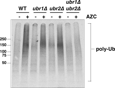
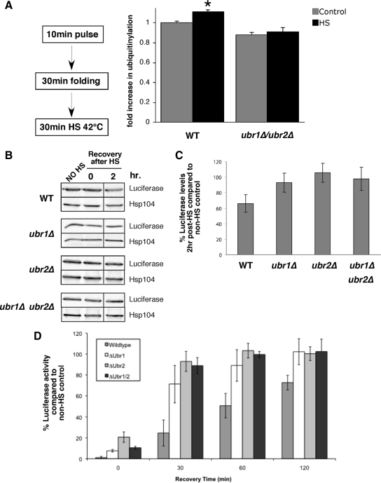
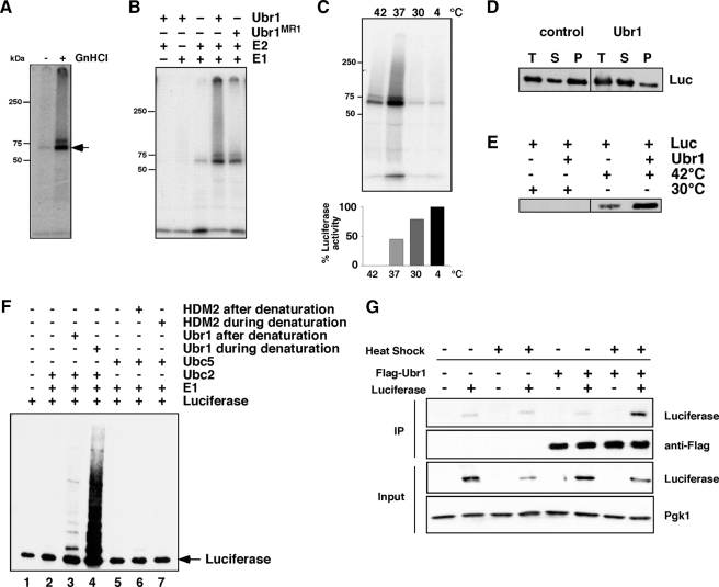
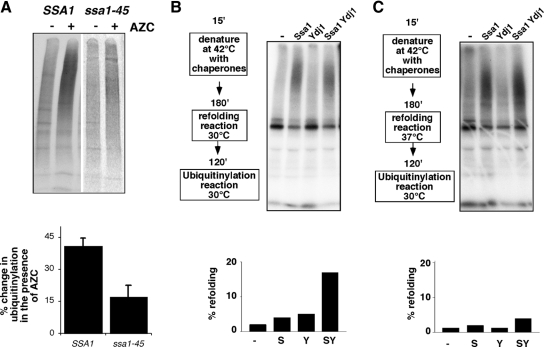
Quality control systems facilitate polypeptide folding and degradation to maintain protein homeostasis. Molecular chaperones promote folding, whereas the ubiquitin/proteasome system mediates degradation. We show here that Saccharomyces cerevisiae Ubr1 and Ubr2 ubiquitin ligases promote degradation of unfolded or misfolded cytosolic polypeptides. Ubr1 also catalyzes ubiquitinylation of denatured but not native luciferase in a purified system. This activity is based on the direct interaction of denatured luciferase with Ubr1, although Hsp70 stimulates polyubiquitinylation of the denatured substrate. We also report that loss of Ubr1 and Ubr2 function suppressed the growth arrest phenotype resulting from chaperone mutation. This correlates with increased protein kinase maturation and indicates partitioning of foldable conformers toward the proteasome. Our findings, based on the efficiency of this quality control system, suggest that the cell trades growth potential to avert the potential toxicity associated with accumulation of unfolded or misfolded proteins. Ubr1 and Ubr2 therefore represent E3 components of a novel quality control pathway for proteins synthesized on cytosolic ribosomes.


Figures

Figure 1.
Figure 1.
Ubr1 functions in the degradation of newly synthesized protein kinases when Hsp90 is inhibited with geldanamycin. (A) Pulse-chase analysis of HA-Tpk2 in wild-type cells treated with geldanamycin (GA; 50 μM) and the proteasome inhibitor, MG132 (100 μM) or solvent alone. (B) Pulse-chase analysis of HA-tagged Tpk2 in wild-type and ubr1Δ cells treated with GA. Chase times shown in minutes. (C) Graph of HA-Tpk2 degradation (n = 3; error bars, ±SE).
Figure 2.
Figure 2.
Ubr1 and Ubr2 promote protein kinase degradation in a cdc37S14A mutant. (A) Pulse-chase analysis of Tap-tagged Tpk2 in wild-type (WT), cdc37S14A (cdc37) mutant cells in the presence and absence of UBR1 (ubr1Δ) and UBR2 (ubr2Δ). 0 and 1 represent times of chase after a 10-min pulse-labeling with [35S]methionine. M, mature and I, immature forms of Tap-tagged Tpk2. (B) Tpk2 activity was measured after IP of Tap-tagged Tpk2 from cell lysates. Assays performed using PepTag nonradioactive detection system. Lysates were from cdc37S14A (cdc37) with and without UBR1 and/or UBR2 as indicated. Results presented as percentage of activity in a wild-type strain. Statistical significance, **p = < 0.01; n = 8. (C) Effect of the proteasome inhibitor, MG132, on Tap-tagged Tpk2 maturation. Chase time shown in minutes. M, mature and I, immature form of the kinase. The relative amount of immature and mature forms are indicated beneath the panel. (D) Assay of yeast cell growth. Tenfold serial dilutions were plated onto YPD plates and incubated at 30 or 34°C as indicated for 3 d. (E) Pulse-chase analysis of Ste11ΔNK444R. Chase times indicated in minutes. Graph at right shows combined results from four independent experiments. Bars, ±SE.
Figure 3.
Figure 3.
Ubr1 functions in protein kinase degradation in association with Rad6. (A) Pulse and 10-min chase analysis of HA-Tpk2 in wild-type (WT) and mutant yeast strains as indicated in the absence and presence of 50 μM GA. Quantitation of band intensity shown in bar graph below panel. (B) As in A., except that chase period was 1 h. Data are from three independent experiments (*p < 0.05; **p < 0.01). (C) Growth of strains in 10-fold serial dilution at 30 and 34°C on YPD plates as indicated.
Figure 4.
Figure 4.
Ubr1 functions in protein kinase degradation independently of the N-end rule. (A) Analysis of Cup9 and Tpk2-TAP degradation in the absence and presence of dipeptides Arg-Ala and Leu-Ala (10 mM each). Pulse-chase analysis shown at the indicated times in the presence of 50 μM GA. (B) As in A, except that the N-end rule substrate, Arg-ß-Gal was analyzed in comparison with HA-Tpk2 in the presence of GA (50 μM) and Arg-Ala/Leu-Ala dipeptides.
Figure 5.
Figure 5.
Ubiquitinylation of newly synthesized polypeptides in the presence of AZC requires Ubr1 and Ubr2. Analysis of ubiquitinylation of newly synthesized polypeptides after a 10-min pulse-labeling in the absence (−) and presence (+) of 50 mM AZC. Polyubiquitinylated polypeptides (poly-Ub) were immunoprecipitated with anti-myc and resolved in a 4–20% denaturing gel before phosphorimaging. Strains used were wild-type (WT), ubr1Δ, ubr2Δ, and ubr1Δ/ubr2Δ double mutants.
Figure 6.
Figure 6.
Ubr1 and Ubr2 promote ubiquitinylation and degradation of mature proteins after heat stress. (A) Wild-type (WT) and ubr1Δ/ubr2Δ cells were pulse-labeled for 10 min and chased with cycloheximide and cold methionine for 30 min. Cells were then incubated at 30 (control) or 42°C (HS) for 30 min. 35S-labeled ubuiquitinylated proteins were immunoprecipitated with anti-Myc and resolved on denaturing gels. The relative amount of ubiquitinylation (fold increase after heat shock) is shown in the bar graph (n = 5; *p = 0.014). (B) Luciferase was expressed from a galactose-inducible promoter in WT, ubr1Δ, ubr2Δ, and ubr/ubr2Δ strains for 2 h. Samples were treated with 0.5 mg/ml cycloheximide, heat shocked (HS) for 60 min at 42°C, and then returned to 30°C for a recovery period. Total protein levels (B and C) of luciferase were monitored before HS, directly after heat shock, and at 2 h after heat shock. Hsp104 is shown as a loading control. (D) Luciferase activity was monitored at the same time points (as described in Materials and Methods). Activity is given as a percent of no heat shock control within each strain background. The data in C and D are plotted as means of three independent experiments. The Western blot shown in B is a representative of the averaged experiments.
Figure 7.
Figure 7.
Binding and ubiquitinylation of denatured luciferase by Ubr1. (A) Purified luciferase was kept native or denatured with guanidinium hydrochloride (GnHCl) before an ubiquitinylation reaction with Ubr1, Ube1, and UbcH2 and 32P-labeled ubiquitin. Reactions were immunoprecipitated with anti-luciferase and were resolved on a denaturing gel before being visualized by autoradiography. Arrow denotes monoubiquitinylated luciferase. (B) Denatured luciferase was incubated with different components of the ubiquitinylation reaction as indicated. A RING mutant of Ubr1, MR1, was also used in this experiment. (C) Purified luciferase was preincubated at the indicated temperatures for 30 min before being split into two aliquots. One was added to an ubiquitinylation reaction containing Ubr1, Ube1 (E1), UbcH2 (E2), and 32P-labeled ubiquitin (top panel). The second aliquot was used to measure remaining luciferase activity (graph in bottom panel). The bars represent luciferase activity as a percentage of the aliquot maintained at 4°C. (D) Purified luciferase was incubated with or without Ubr1 before denaturation at 42°C. The total reaction (T) was separated into supernatant (S) and pellet (P) fractions after centrifugation. The amount of luciferase in each fraction was determined by Western blot. (E) Luciferase was kept native or denatured at 42°C in the absence or presence of Ubr1. Ubr1 was immunoprecipitated, and the amount of luciferase that coimmunoprecipitated was determined by Western blot. (F) Ubiquitinylation of luciferase when Ubr1 is present during the denaturation step. Luciferase was denatured by incubation at 42°C by itself (lane 1) or in the presence of Ubr1 (lane 4) or HDM2 (lane 7). The luciferase was then added to reactions containing E1 and Ubc2 (lane 2); E1, Ubc2, and Ubr1 (lane 3); E1 and Ubc5 (lane 5); and E1, Ubc5, and HDM2 (lane 6). All reactions contained ATP and ubiquitin. Luciferase was visualized by Western blot. Arrow denotes nonubiquitinylated luciferase. (G) IP of Flag-Ubr1 and Western blot for coimmunoprecipitating luciferase before and after heat shock at 42°C for 30 min.
Figure 8.
Figure 8.
Hsp70 promotes ubiquitinylation by Ubr1. (A) Ubiquitinylation of newly synthesized polypeptides in the absence (−) and presence (+) of AZC in strains expressing myc-tagged ubiquitin. IP of 35S-labeled polypeptides with anti-myc is shown. Strains used were wild type (JN516; SSA1) and ssa1-45. Bar graph below panel indicates levels of induction of ubiquitinylated newly synthesized polypeptides. Bars, SE; n = 3. (B) Ubiquitinylation reaction of heat-denatured luciferase using 32P-labeled ubiquitin. Purified Ssa1 (5:1 with luciferase) or Ydj1 (15:1 with luciferase) were added to the heat denaturation reaction, followed by a further incubation (“chaperone incubation”) and the subsequent ubiquitinylation reaction using 32P-labeled ubiquitin as indicated. In this reaction, the denaturation was followed by chaperone and ubiquitinylation reactions both at 30°C. The reaction was split into two before the ubiquitinylation reaction. One-half was used for the ubiquitinylation reaction, and the second half used for assays of luciferase activity, shown in the graph below the panel. (C) Same as B, except that the chaperone reaction was performed at 37°C.

Similar articles

Cited by

References

    1. Arndt V., Rogon C., Hohfeld J. To be, or not to be—molecular chaperones in protein degradation. Cell Mol. Life Sci. 2007;64:2525–2541. - PMC - PubMed
    1. Baker R. T., Varshavsky A. Inhibition of the N-end rule pathway in living cells. Proc. Natl. Acad. Sci. USA. 1991;88:1090–1094. - PMC - PubMed
    1. Balch W. E., Morimoto R. I., Dillin A., Kelly J. W. Adapting proteostasis for disease intervention. Science. 2008;319:916–919. - PubMed
    1. Bartel B., Wunning I., Varshavsky A. The recognition component of the N-end rule pathway. EMBO J. 1990;9:3179–3189. - PMC - PubMed
    1. Becker J., Walter W., Yan W., Craig E. A. Functional interaction of cytosolic hsp70 and a Dnaj-related protein, Ydj1p, in protein translocation in vivo. Mol. Cell Biol. 1996;16:4378–4386. - PMC - PubMed

Publication types

MeSH terms