. 2010 Jul 1;21(13):2315-26.
doi: 10.1091/mbc.e09-11-0953. Epub 2010 May 12.

Distinct contributions of conserved modules to Runt transcription factor activity

Affiliations

Distinct contributions of conserved modules to Runt transcription factor activity

Pegine B Walrad et al. Mol Biol Cell. .

Abstract

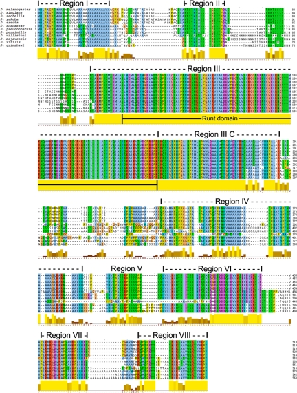
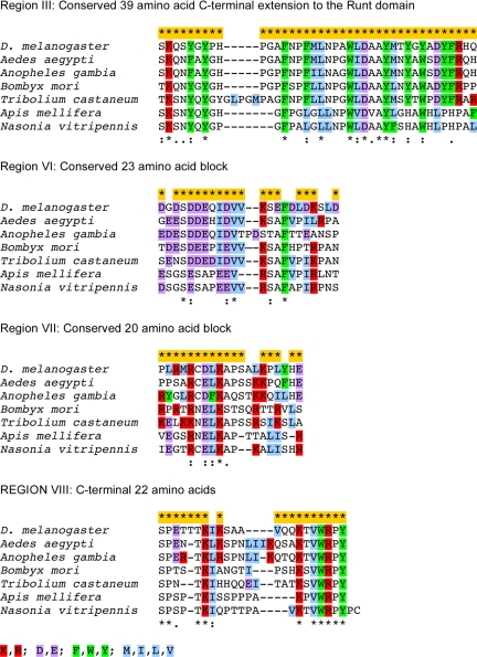
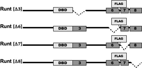
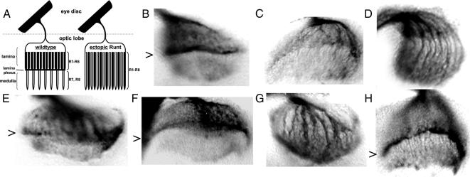
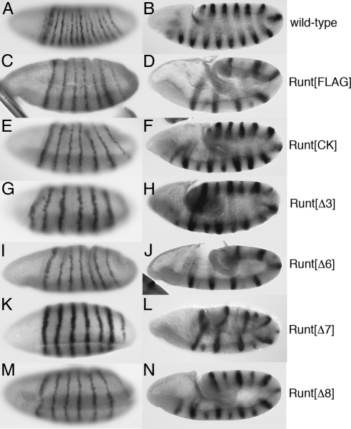
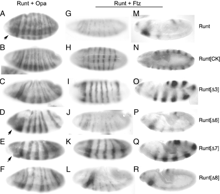
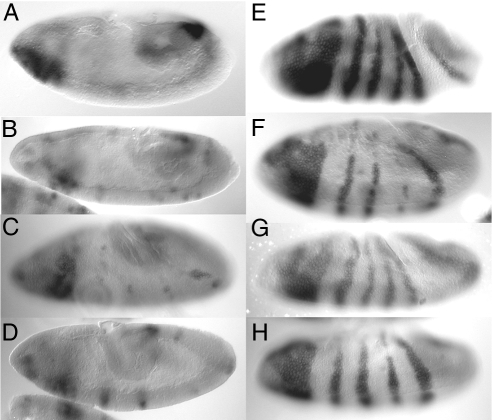
Runx proteins play vital roles in regulating transcription in numerous developmental pathways throughout the animal kingdom. Two Runx protein hallmarks are the DNA-binding Runt domain and a C-terminal VWRPY motif that mediates interaction with TLE/Gro corepressor proteins. A phylogenetic analysis of Runt, the founding Runx family member, identifies four distinct regions C-terminal to the Runt domain that are conserved in Drosophila and other insects. We used a series of previously described ectopic expression assays to investigate the functions of these different conserved regions in regulating gene expression during embryogenesis and in controlling axonal projections in the developing eye. The results indicate each conserved region is required for a different subset of activities and identify distinct regions that participate in the transcriptional activation and repression of the segmentation gene sloppy-paired-1 (slp1). Interestingly, the C-terminal VWRPY-containing region is not required for repression but instead plays a role in slp1 activation. Genetic experiments indicating that Groucho (Gro) does not participate in slp1 regulation further suggest that Runt's conserved C-terminus interacts with other factors to promote transcriptional activation. These results provide a foundation for further studies on the molecular interactions that contribute to the context-dependent properties of Runx proteins as developmental regulators.


Figures

Figure 1.
Figure 1.
Conservation of the Runt protein in Drosophila. The figure shows a ClustalW2-generated alignment of Runt protein sequences from 12 different Drosophila species. The D. melanogaster amino acid sequence (single -letter code) is given at the top of each segment of the alignment with the other species listed in the order of their increasing divergence from D. melanogaster. The top five species (D. melanogaster, D. simulans, D. sechellia, D. yakuba and D. erecta) comprise the melanogaster subgroup. The melanogaster group includes these five plus D. ananassae. The color-coding of conserved regions in the alignment is as provided by ClustalW2: Hydrophobic (A, F, L, M, V, W), light blue; Basic (K, R), red; Acidic (D, E), purple; Polar (N, Q, S, T), green; C, pink; G, salmon; and H and Y, blue. The limits of the eight conserved regions identified in the initial three-way alignment are indicated above the D. melanogaster sequence. These initial limits were used to guide the generation of the deletion constructs used to investigate the in vivo functions of the different conserved regions. A ClustalW2-generated plot of sequence conservation is provided across the bottom for each of the different sequence segments. Positions that are conserved with sequence identity in all species are indicated in yellow in this plot, with an asterisk (*) below the amino acid position. The limits of the Runt domain are indicated within the extended block of sequence conservation revealed in the plot for region III. The region I alignment shown in the figure fails to identify a conserved pentapeptide motif (S/T)QVL(Q/A) that precedes a homopolymeric run of eight (D. willistoni) to 12 (all of the others except D. ananassae, D. psuedoobscura, and D. persimilis) alanine residues.
Figure 2.
Figure 2.
Conservation of RUNT C-terminal modules in other insects. The figure shows an alignment of the regions of Runt proteins from six nondrosophilid insects with corresponding intervals from conserved regions III, VI, VII, and VIII of the Drosophila proteins. The top line of sequence information in each segment is from D. melanogaster, with residues that are conserved with identity in all 12 Drosophila species indicated by a yellow bar and an asterisk (*) above the sequence. Species identification is provided to the left of each of the other sequence segments. Residues that are conserved with identity in all of the sequences in this alignment are indicated below the alignment with an asterisk, conserved substitutions are indicated with a colon, and similarities are indicated with a period. A legend for the color coding used to identify basic, acidic, aromatic, and hydrophobic amino acids is provided at the bottom of the figure.
Figure 3.
Figure 3.
Runt deletion constructs. Schematic diagram indicating the regions removed in different UAS-Runt deletion constructs and the location of the FLAG epitope tag between conserved regions VI and VII. The solid horizontal line represents the Runt protein, with boxed regions on this line indicating the relative locations of different conserved regions. Regions removed in different deletion constructs are indicated by dashed lines that connect the regions flanking the deletions. Runt[Δ3] removes the segment of conserved region III that is immediately C-terminal to the Runt domain (DBD).
Figure 4.
Figure 4.
Expression and nuclear localization of the Runt deletion derivatives. Expression of different Runt deletion constructs in larval salivary glands as detected by the anti-FLAG M2 mAb. (A–F) Background antibody control of salivary glands that are not expressing a FLAG-tagged protein (A), FLAG-tagged full-length Runt (B), Runt[Δ3] (C), Runt[Δ6] (D), Runt[Δ7] (E), and Runt[Δ8] (F). The images in A, C, and E have higher background fluorescence due to differences in the antibody used for detection and the imaging instrumentation when the experiment was extended to include Runt[Δ3]. Images for Runt[Δ7] were acquired using both sets of conditions and indicate the difference is due to background, and not differences in expression levels of the Runt deletion derivatives. The images with higher background are used for the control in A and the Runt[Δ3] and Runt[Δ7] proteins as this best demonstrates their similarity in this assay.
Figure 5.
Figure 5.
Functional specificity of Runt-dependent axonal pathfinding. (A) Schematic of eye disk and optic lobe in Drosophila third-instar larvae. Axons of wild-type photoreceptor neurons R1–R6 terminate between the lamina and medulla of the optic lobe forming the lamina plexus. Axons of photoreceptors R7 and R8 that normally express Runt terminate in the medulla. (B) Optic lobe of wild-type larvae with axonal projections revealed by immunodetection of photoreceptor membrane-associated chaoptin. The arrowhead indicates the lamina plexus. (C) MT14 GAL4-driven UAS-Runt expression in R2 and R5 redirects all axons to terminate in the medulla leading to elimination of the lamina plexus. (D–H) Axonal projections in optic lobes of larvae with MT14 driven expression of UAS-Runt[CK]77 (D), UAS-Runt[Δ3]46-1 (E), UAS-Runt[Δ6]3-1 (F), UAS-Runt[Δ7]45-2 (G), and UAS-Runt[Δ8]49 (H), with arrowheads indicating preparations that retain a lamina plexus.
Figure 6.
Figure 6.
Differential requirements in establishing and maintaining en repression. Expression of en mRNA as revealed by in situ hybridization in gastrula stage (left column) and germband extension stage (right column) Drosophila embryos. (A and B) Wild-type expression at these two stages. (C–N) The response of en to NGT-driven expression of UAS-Runt[FLAG]1-3 (C and D), UAS-Runt[CK]77 (E and F), UAS-Runt[Δ3]46-1 (G and H), UAS-Runt[Δ6]3-1 (I and J), UAS-Runt[Δ7]45-2 (K and L), and UAS-Runt[Δ8]49 (M and N). Embryos with ectopic Runt expression were generated by mating homozygous NGT40 females to males carrying the pertinent UAS-Runt transgene for all crosses except for those with the Runt[Δ8], which were done with homozygous NGT40 + NGTA females, which produce ∼1.5× the level of NGT-driven expression as NGT40. The three Runt derivatives that maintain en repression, Runt[FLAG], Runt[Δ6] and Runt[Δ7], all give completely penetrant repression in gastrula and early germband extension stage embryos that is stably maintained in 45 of 46 stage 10 and 11 embryos with Runt[FLAG], 13 of 26 embryos generated in crosses with UAS-Runt[Δ6]3-1/TM3 males, and in 19 of 21 Runt[Δ7]-expressing embryos. The inability of Runt[CK] and Runt[Δ8] to maintain en repression has been described previously (Wheeler et al., 2002). A similar loss of this activity is observed for Runt[Δ3], which in this experiment produced fully penetrant early repression (29 of 29 gastrula and early germband extension stage embryos) that was not maintained later (43 of 43 stage 10 and 11 embryos express the odd-numbered en stripes, with 20 of 43 showing equal levels of expression of even and odd stripes). The de-repression of odd-numbered en stripes in these later stages is also observed for the Runt[CK], Runt[Δ3], and Runt[Δ8] (as shown in N) at the higher ectopic expression levels obtained using homozygous NGT40 + NGTA females.
Figure 7.
Figure 7.
Differential requirements for slp1 activation and repression. Expression of slp1 mRNA as revealed by in situ hybridization. (A–F) gastrula stage expression of slp1 in response to NGT-driven coexpression of Opa and different Runt deletion derivatives. In all cases ectopic expression was obtained using females homozygous for both the NGT40 and NGTA GAL4 drivers. (A) UAS-Runt232 and UAS-Opa12, 60 of 65 gastrula stage embryos scored in this experiment showed ectopic anterior slp1 activation comparable or stronger than that shown in this panel. The remaining five embryos had weaker anterior activation with incomplete fusion of stripes within the segmented region of the embryo. (B) UAS-Runt[CK]77 and UAS-Opa12, 32 of 39 gastrula stage embryos showed incomplete fusion of slp1 stripes similar to that depicted in this panel, and six of seven the remaining embryos showed evidence of weak anterior activation, with one embryo showing clear evidence of ectopic anterior activation. (C) UAS-Runt[Δ3]46-1 and UAS-Opa14, 34 of 37 embryos showed incomplete fusion as depicted, with the other three showing evidence of weak anterior activation. (D) UAS-Runt[Δ6]3-1/TM3 and UAS-Opa14, 13 of 25 gastrula stage embryos showed strong anterior activation similar to that shown in this panel. As expected in a cross with males heterozygous for the UAS-Runt[Δ6] construct, 12 of 25 showed minor alterations in the spacing of slp1 stripes produced by NGT-driven expression of Opa alone. (E) UAS-Runt[Δ7]21-3/TM3 and UAS-Opa14, 13 of 28 gastrula stage embryos in crosses with these heterozygous males showed strong anterior activation. (F) UAS-Runt[Δ8]49 and UAS-Opa10, 28 of 38 gastrula stage embryos showed abnormal spacing of slp1 stripes, whereas 10 of 38 showed loss of specific stripes similar to that shown, presumably due to repression by Runt[Δ8] (see below). None of the embryos in this cross showed strong anterior activation. Arrows indicate regions of anterior slp1 activation in response to Runt, Runt[Δ6], and Runt[Δ7]. The potent activity of Runt[Δ7] in slp1 activation is underscored by the use of the weaker UAS-Runt[Δ7]21-3 line in this coexpression assay. Similarly, the inability of the Runt[Δ8] derivative to activate slp1 is underscored by the use of UAS-Opa10 as this line is stronger than the UAS-Opa lines used for the other Runt constructs (Swantek and Gergen, 2004). The slp1 response to NGT-driven coexpression of Ftz and these different Runt deletion derivatives in gastrula (G–L) and germband extension stages (M–R). In all cases ectopic expression was obtained by mating females homozygous for both NGT40 and NGTA to males homozygous for UAS-Ftz263 and the pertinent Runt transgene: (G and M) UAS-Runt232, 15 of 22 gastrula stage embryos show partial to complete repression of the even-numbered slp1 stripes; (H and N) UAS-Runt[CK]77, 0 of 23 gastrula stage embryos showed any evidence of repression of the even-numbered stripes; (I and O) UAS-Runt[Δ3]46-1, 0 of 8 gastrula stage embryos show repression of all of the even-numbered stripes, although there is a region specific reduction in stripe 10 expression in several embryos; (J and P) UAS-Runt[Δ6]16-2, 10 of 10 gastrula stage embryos show evidence of repression¤ with four of these having nearly complete repression similar to that shown in this panel. Note that the Runt[Δ6] line used in this experiment is slightly weaker than the line used in the slp1 activation assay described above; (K and Q) UAS-Runt[Δ7]45-2, 0 of 17 gastrula stage embryos show repression of all even-numbered stripes, although expression of stripe 10 is reduced in 16 of these 17 embryos; (L and R) UAS-Runt[Δ8]49/TM3, 17 of 33 gastrula and early germband extension stage embryos scored in the cross with these heterozygous males showed partial to complete repression of the even-numbered stripes. The odd-numbered slp1 stripes are repressed in all of the embryos in these different crosses due to ectopic Ftz expression in cells expressing endogenous Runt.
Figure 8.
Figure 8.
Runt-dependent slp1 regulation is insensitive to Gro and Rpd3 dosage. Expression of slp1 mRNA in response to NGT-driven coexpression of UAS-Runt15 and UAS-Ftz263 (A–D) or UAS-Runt15 and UAS-Opa14 (E–H) transgenes. (A) Sixteen of 20 gastrula and early germband extension stage embryos from crosses involving females heterozygous for both the NGT40 and NGTA drivers that are wild-type for Gro and Rpd3 show partial to complete repression of slp1. Reducing the strength of either the NGT driver or the UAS-Runt line reduces the efficiency of the repression of slp1 observed in response to coexpression of Runt and Ftz (Swantek and Gergen, 2004). (B) Nineteen of 23 embryos from females heterozygous for NT40, NGTA, and the GroBX22 mutation show repression of slp1. (C) Nineteen of 25 embryos from heterozygous NGT40, NGTA females that are also heterozygous for GroE48 show slp1 repression. (D) Thirty of 36 embryos from females heterozygous for NGT40, NGTA, and the Rpd304556 mutation show evidence of repression. (E) Anterior activation in response to of Runt and Opa is observed in 57 of 58 embryos from females heterozygous for NGT40 and NGTA that are otherwise wild type. Strong anterior activation is also observed in crosses with females that are also heterozygous for (F) GroBX22, 20 of 23 embryos; (G) GroE48, 30 of 39 embryos; and (H) Rpd304556, 46 of 55 gastrula and early germband extension stage embryos.

Similar articles

Cited by

References

    1. Aronson B. D., Fisher A. L., Blechman K., Caudy M., Gergen J. P. Groucho-dependent and -independent repression activities of Runt domain proteins. Mol. Cell. Biol. 1997;17:5581–5587. - PMC - PubMed
    1. Bao R., Friedrich M. Conserved cluster organization of insect Runx genes. Dev. Genes Evol. 2008;218:567–574. - PubMed
    1. Bravo J., Li Z., Speck N. A., Warren A. J. The leukemia-associated AML1 (Runx1)–CBF beta complex functions as a DNA-induced molecular clamp. Nat. Struct. Biol. 2001;8:371–378. - PubMed
    1. Damen W.G.M., Weller M., Tautz D. Expression patterns of hairy, even-skipped and runt in the spider Cupiennius salei imply that these genes were segmentation genes in a basal arthropod. Proc. Natl. Acad. Sci. USA. 2000;97:4515–4519. - PMC - PubMed
    1. de Bruijn M. F., Speck N. A. Core-binding factors in hematopoiesis and immune function. Oncogene. 2004;23:4238–4248. - PubMed

Publication types

MeSH terms

Substances

LinkOut - more resources