. 2010 Jul 1;21(13):2285-96.
doi: 10.1091/mbc.e09-11-0947. Epub 2010 May 12.

TBC-2 regulates RAB-5/RAB-7-mediated endosomal trafficking in Caenorhabditis elegans

Affiliations

TBC-2 regulates RAB-5/RAB-7-mediated endosomal trafficking in Caenorhabditis elegans

Laëtitia Chotard et al. Mol Biol Cell. .

Abstract

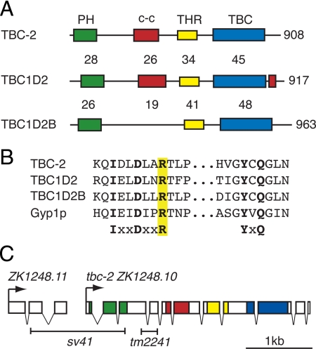
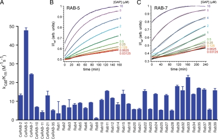
During endosome maturation the early endosomal Rab5 GTPase is replaced with the late endosomal Rab7 GTPase. It has been proposed that active Rab5 can recruit and activate Rab7, which in turn could inactivate and remove Rab5. However, many of the Rab5 and Rab7 regulators that mediate endosome maturation are not known. Here, we identify Caenorhabditis elegans TBC-2, a conserved putative Rab GTPase-activating protein (GAP), as a regulator of endosome to lysosome trafficking in several tissues. We show that tbc-2 mutant animals accumulate enormous RAB-7-positive late endosomes in the intestine containing refractile material. RAB-5, RAB-7, and components of the homotypic fusion and vacuole protein sorting (HOPS) complex, a RAB-7 effector/putative guanine nucleotide exchange factor (GEF), are required for the tbc-2(-) intestinal phenotype. Expression of activated RAB-5 Q78L in the intestine phenocopies the tbc-2(-) large late endosome phenotype in a RAB-7 and HOPS complex-dependent manner. TBC-2 requires the catalytic arginine-finger for function in vivo and displays the strongest GAP activity on RAB-5 in vitro. However, TBC-2 colocalizes primarily with RAB-7 on late endosomes and requires RAB-7 for membrane localization. Our data suggest that TBC-2 functions on late endosomes to inactivate RAB-5 during endosome maturation.


Figures

Figure 1.
Figure 1.
TBC-2: protein homology, gene structure, and deletion alleles. (A) C. elegans TBC-2 (NP_495156) is homologous to the human TBC1D2 (Q9BYX2) and TBC1D2B (Q9UPU7) proteins. They share a conserved PH domain (green), coiled-coil domain (c-c, red) and TBC domain (blue). The homology between TBC-2 and its mammalian homologues to mainly limited to the three domains with the exception of a region we designated the THR (yellow). The percentage of amino acid identity between TBC-2 and the two human homologues is shown above each domain. (B) Alignment of the TBC catalytic motifs IXXDXXR and YXQ containing the conserved arginine (R; highlighted yellow) and glutamine (Q) residues conserved in C. elegans TBC-2, human TBC1D2, and TBC1D2B and yeast Gyp1p (NP_014713). (C) Genomic structure of tbc-2 ZK1248.10 and the upstream gene ZK1248.11. The regions encoding the different domains in TBC-2 are highlighted in corresponding colors. Shown below are the regions deleted by the deletion alleles sv41 and tm2241. The sv41 allele removes most of ZK1248.11 in addition to the 5′ region of tbc-2. The tm2241 allele removes intron three resulting in a frame shift introducing a premature stop codon.
Figure 2.
Figure 2.
tbc-2() phenotypes in the intestine, coelomocytes, and oocytes. (A and B) DIC images of wild-type (A) and tbc-2(tm2241) (B) animals. The intestine is composed of 20 polarized epithelial cells with an apical membrane facing the intestinal lumen (arrow) and basolateral membranes facing the pseudocoelomic space and neighboring organs (boundaries marked with bracket). Examples of the large vesicles observed in tbc-2() animals are marked with arrowheads. (C–F) Electron micrographs of transverse sections of wild-type (C) and tbc-2(tm2241) (D–F) animals. (C and D) A white line demarcates the basolateral boundaries of the intestine (int) from other tissues such as the gonad (go), and the intestinal lumen is marked with an arrow. (E and F) Higher magnification images of the large tbc-2(tm2241) vesicles and the amorphous material present inside the vesicles. (G and H) Epifluorescence images of coelomocytes containing internalized ssGFP in arIs37[Pmyo-3::GFP]; dpy-20(e1282) (G) and tbc-2(tm2241); arIs37[Pmyo-3::GFP] (H) animals. (I) Scatter dot plot of the largest ssGFP-positive vesicle in each of 60 coelomocytes from arIs37[Pmyo-3::GFP]; dpy-20(e1282) (wild-type) and tbc-2(tm2241); arIs37[Pmyo-3::GFP] animals. The difference between the two groups is statistically significant in an unpaired t test (p < 0.0001). Error bars represent the mean with a 95% confidence interval (p < 0.05). (J–M) DIC (J and L) and epifluorescence (K and M) images of oocytes with internalized YP170::GFP in bIs1[vit-2::GFP] (J and K) and tbc-2(tm2241); bIs1[vit-2::GFP] (L and M) animals. Bar, 10 μm (A, B, G, H, J, K, L, and M), 5 μm (C and D), 0.5 μm (E) and 0.2 μm (F).
Figure 3.
Figure 3.
tbc-2() intestinal vesicles are large endosomes. (A–P) Confocal images of wild-type (A, C, E, G, I, K, M, and O) and tbc-2(tm2241) (B, D, F, H, J, L, N, and P) animals carrying the following integrated transgenes: pwIs72 [Pvha-6::GFP::rab-5] (A and B), pwIs170 [Pvha-6::GFP::rab-7] (C and D), pwIs50 [lmp-1::GFP] (E and F), pwIs50 [lmp-1::GFP] and pwIs429[Pvha-6::mCherry::rab-7] (G and H), pwIs50 [lmp-1::GFP] fed LysoTracker Red (red) (I and J), pwIs206 [Pvha-6::GFP::rab-10] (K and L), pwIs69 [Pvha-6::GFP::rab-11] (M and N), and an extrachromosomal array carrying GFP::lgg-1 (Melendez et al., 2003) (O and P). Arrowheads denote examples of GFP::RAB-5–positive vesicles (B), vesicles with internalized GFP::RAB-7 (D), LMP-1::GFP (F), or GFP::LGG-1 (P), and LysoTracker Red-positive vesicles adjacent to LMP-1::GFP vesicles (J). Bar, 5 μm.
Figure 4.
Figure 4.
TBC-2 rescue and localization. Confocal images of tbc-2(tm2241) animals expressing either vhIs1[Pvha-6::mCherry::tbc-2] (A–C, G–I, M–O, and S–U), or vhIs6[Pvha-6::mCherry::tbc-2 R689K] (D–F, J–L, P–R, and V–X) in combination with pwIs72[Pvha-6::GFP::rab-5] (A–F), pwIs170[Pvha-6:: GFP::rab-7] (G–L), pwIs50[lmp-1::GFP] (M-R), pwIs69[Pvha-6::GFP::rab-11] (S–X). Bar, 5 μm.
Figure 5.
Figure 5.
rab-5, rab-7, and the HOPS complex are required for tbc-2() intestinal phenotype. (A–J) DIC images (A, C, E, G, and I) and epifluorescence images (B, D, F, H, and J) of the intestines of tbc-2(tm2241) animals expressing LMP-1::GFP in different mutant and RNAi backgrounds. (A and B) tbc-2(tm2241) unc-4(e120); rde-1(ne219); pwIs50 [lmp-1::GFP]; rrEx236[Pend-3::rde-1 + Pelt-2::rde-1 + Pinx-6::GFP] animals fed dsRNA corresponding to rab-5. This strain was used for intestinal specific RNAi (see Materials and Methods). (C and D) tbc-2(tm2241) rab-7(ok511); pwIs50 [lmp-1::GFP] animals. (E–J) tbc-2(tm2241); pwIs50 [lmp-1::GFP] fed dsRNA corresponding to rab-11.1 (E and F), vps-39 (G and H), or vps-41 (I and J). Confocal images of pwIs170[Pvha-6::GFP::rab-7] (K and L), and tbc-2(tm2241); pwIs170[Pvha-6::GFP::rab-7] (M and N) animals fed dsRNA targeting vps-39. (L and N) are higher magnification images from K and M, respectively. Bar, 10 μm (B and K) and 2 μm (L).
Figure 6.
Figure 6.
rab-7 is required for the YP170::GFP distribution in tbc-2() oocytes. DIC (A, C, and E) and epifluorescence (B, D, and F) images of oocytes of tbc-2(tm2241) (A and B), rab-7(RNAi) (C and D) and tbc-2(tm2241) rab-7(RNAi) (E and F) animals expressing YP170::GFP. Bar, 10 μm (D).
Figure 7.
Figure 7.
Expression of RAB-5 Q78L mimics the tbc-2() intestinal phenotype in a RAB-7 and HOPS complex dependent manner. (A–D) DIC images (A and C) and epifluorescence images (B and D) of the intestine of vhIs24 [Pvha-6::GFP::rab-5 Q78L] (A and B) and vhIs34[Pvha-6::GFP::rab-7 Q68L] (C and D) transgenic animals. (E–G) Confocal images of the intestine of a strain expressing both vhIs24 [Pvha-6::GFP::rab-5 Q78L] and pwIs429[Pvha-6:: mCherry::RAB-7]. Arrowheads denote smaller vesicles positive for both GFP::RAB-5 Q78L and mCherry::RAB-7, and asterisks denotes larger mCherry::RAB-7–positive vesicles almost devoid of GFP::RAB-5 Q78L. (H–K) DIC images of the intestine of vhIs24 [Pvha-6:: GFP::rab-5 Q78L] transgenic animals fed with dsRNA targeting GFP (H), rab-7 (I), vps-39 (J), or vps-41 (K). Bar, 10 μm (D and K) and 5 μm (G).
Figure 8.
Figure 8.
TBC-2 catalyzes GTP hydrolysis by RAB-5 in vitro. (A) Catalytic efficiency (kcat/Km) plot based on the initial velocity measurements of TBC-2 on the C. elegans (Ce) and mammalian Rab GTPases. (B and C) A global fitting analysis to a Michaelis–Menten model function (in the limit where [TBC-2] < Km) for RAB-5 (B) and RAB-7 (C) at different concentrations of TBC-2. Solid lines represent the fitted model functions.
Figure 9.
Figure 9.
RAB-7 is required for TBC-2 localization to membranes. Confocal images of tbc-2(tm2241); vhIs12 [Pvha-6::GFP::tbc-2] animals fed an empty vector (A and B) or dsRNA targeting rab-7 (C and D). B and D are higher magnification images from A and C, respectively. Experiments were performed in the tbc-2(tm2241) background as GFP::TBC-2 membrane localization was more robust, presumably due to competition with endogenous TBC-2. Bar, 10 μm (A) and 2 μm (B).

Similar articles

Cited by

References

    1. Albert S., Gallwitz D. Two new members of a family of Ypt/Rab GTPase activating proteins. Promiscuity of substrate recognition. J. Biol. Chem. 1999;274:33186–33189. - PubMed
    1. Albert S., Will E., Gallwitz D. Identification of the catalytic domains and their functionally critical arginine residues of two yeast GTPase-activating proteins specific for Ypt/Rab transport GTPases. EMBO J. 1999;18:5216–5225. - PMC - PubMed
    1. Bernards A. GAPs galore! A survey of putative Ras superfamily GTPase activating proteins in man and Drosophila. Biochim. Biophys. Acta. 2003;1603:47–82. - PubMed
    1. Brenner S. The genetics of Caenorhabditis elegans. Genetics. 1974;77:71–94. - PMC - PubMed
    1. Brune M., Hunter J. L., Corrie J. E., Webb M. R. Direct, real-time measurement of rapid inorganic phosphate release using a novel fluorescent probe and its application to actomyosin subfragment 1 ATPase. Biochemistry. 1994;33:8262–8271. - PubMed

Publication types

MeSH terms

Substances