Review
. 2011 Aug 1;3(8):a003616.
doi: 10.1101/cshperspect.a003616.

Group II introns: mobile ribozymes that invade DNA

Affiliations
Review

Group II introns: mobile ribozymes that invade DNA

Alan M Lambowitz et al. Cold Spring Harb Perspect Biol. .

Abstract

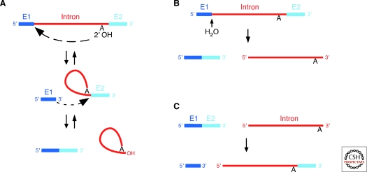
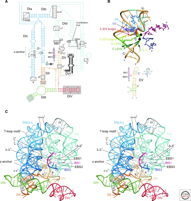
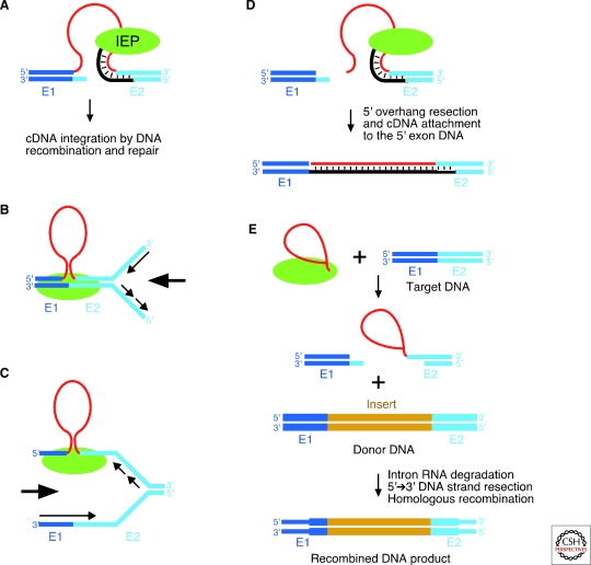
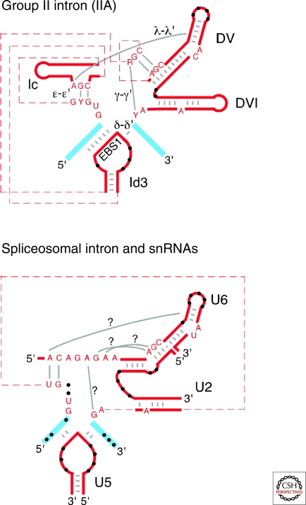
Group II introns are mobile ribozymes that self-splice from precursor RNAs to yield excised intron lariat RNAs, which then invade new genomic DNA sites by reverse splicing. The introns encode a reverse transcriptase that stabilizes the catalytically active RNA structure for forward and reverse splicing, and afterwards converts the integrated intron RNA back into DNA. The characteristics of group II introns suggest that they or their close relatives were evolutionary ancestors of spliceosomal introns, the spliceosome, and retrotransposons in eukaryotes. Further, their ribozyme-based DNA integration mechanism enabled the development of group II introns into gene targeting vectors ("targetrons"), which have the unique feature of readily programmable DNA target specificity.


Figures

Figure 1.
Figure 1.
Group II intron RNA secondary structure. (A) Structure of a representative bacterial IIA1 intron (not to scale), with notable variations in IIB and IIC introns shown in circles. Boxes indicate sequences involved in tertiary interactions (Greek letters, EBS, IBS). The “loop” of DIV, which encodes the IEP, is depicted by dashed lines, with a box showing the location and structure of DIVa of the Lactococcus lactis Ll.LtrB intron, a high-affinity binding site for the IEP. Subdomains discussed in the text are labeled, with base pairs (dashes) shown only for DV and the κ-stem-loop. Compared to IIA introns, major differences in other subgroups include structural features of DV (IIC introns); different ε′ motifs (IIB, IIC); the number of base pairs in the κ-stem-loop (IIC); a coordination loop containing EBS3 and δ′ (IIB, IIC); the absence of the DId(iii) stem-loop (IIB, IIC); the absence of a stem in the EBS2 motif (IIB, IIC); a unique ζ–ζ′ motif (IIC); and the ω–ω′ interaction (IIC, some IIB). (B) Base-pairing interactions used by IIA, IIB, and IIC introns to bind the exons at the active site. EBS, exon-binding site; IBS, intron-binding site.
Figure 2.
Figure 2.
Group II intron IEPs and related proteins. (A) LtrA protein encoded by the L. lactis Ll.LtrB intron. (B) IEP lacking an En domain encoded by the Sinorhizobium meliloti RmInt1 intron, which belongs to bacterial lineage D (see Fig. 3). The “class D motif” at the carboxy-terminus is a conserved sequence that is required for splicing and mobility functions in lineage D IEPs (Molina-Sánchez et al. 2010). (C) MatK protein encoded by the Arabidopsis thaliana trnKI1 intron. MatK proteins retain conserved sequence blocks RT5-7 and domain X, but their amino-terminal halves have diverged from those of canonical group II IEPs, and they lack an En domain (Mohr et al. 1993). (D) nMat-1a protein encoded by a nuclear gene in Arabidopsis thaliana. nMat-1 proteins contain complete RT and X domains, but have mutations expected to inhibit RT activity; nMat-2 proteins (not shown) also contain an En domain, but with mutations expected to inhibit En activity (Mohr and Lambowitz 2003). (E) LAGLIDADG protein encoded by Cryphonectria parasitica rrnI1. (F) HIV-1 RT. Schematics of introns and ORFs are to scale. Insertions between RT sequence blocks are denoted 2a, 3a, 4a, and 7a. The locations of the three-predicted α-helices characteristic of thumb domains are shown above domain X in LtrA (cf. with HIV-1 RT in panel F).
Figure 3.
Figure 3.
Group II intron lineages. The major lineages of group II intron IEPs, denoted CL (chloroplast-like), ML (mitochondrial-like), and bacterial classes A-F, are shown as blue sectors. Notable sublineages, including four subdivisions of CL and a subclass of IIC introns that inserts after attC sites, are shown as darker blue sectors within the major lineages. RNA structural subgroups that correspond to IEP lineages are shown in magenta. All group II intron lineages and RNA types are found in bacteria. Lineages and RNA types also found in organelles are delineated in green (outer circle). Note that there may be limited exceptions to the overall pattern of co-evolution within the CL group, with different sublineages possibly having exchanged IIB RNA structures (Simon et al. 2009). An alternate nomenclature for group II lineages has been proposed, which does not distinguish between IEP and ribozyme lineages or take into account exceptions to their coevolution (Toro et al. 2002).
Figure 4.
Figure 4.
Reactions catalyzed by group II introns RNAs. (A) Forward and reverse splicing. (B) Hydrolytic splicing. The initial step is hydrolytic cleavage at the 5′ splice site. The second step leading to exon ligation (not shown) is the same as for splicing via lariat formation (panel A). (C) Partial reverse splicing by linear intron RNA, leading to ligation of the 3′ end of the intron RNA to the 5′ end of the 3′ exon. Intron RNA, red; 5′ and 3′ exons (E1 and E2), dark and light blue, respectively.
Figure 5.
Figure 5.
Crystal structure of the Oceanobacillus iheyensis group IIC intron. (A) Sequence and secondary structure of the crystallized RNA. Boxes indicate motifs involved in tertiary interactions. Solid gray boxes in DII, DIII, and DIV indicate regions deleted from the crystallization construct and replaced with sequences not present in the wild-type intron. Nucleotide residues in DI and DVI shown as white letters on a black background are not visible in the crystal structure. (B) Structure of the active-site region, with a corresponding color-coded secondary structure below. DV is a beige tube helix, with a bound RNA modeled as the 5′ and 3′ exons (pink and indigo, respectively; Toor et al. 2008b; Toor et al. 2010). The triple interactions in the triple helix stack between the CGC triad, the CG of J2/3, and the C of the AC bulge are shown in dark green, green and yellow-green. The three-base stack consisting of the A of the AC bulge, G5, and U4 of the ε–ε′ interaction are in red, purple and blue, respectively, while the λ–λ′ interaction is cyan. Metal ions bound to DV in the crystal are indicated by spheres, with black spheres representing the proposed active-site Mg++ ions, which were identified by binding of Yb3+ in the crystal derivatives. (C) X-ray crystal structure. A stereoview is shown, with domains colored as in (A) and regions involved in tertiary interactions colored gray (Toor et al. 2008a,b). Note added in proof: The conserved single base pair in the κ stem-loop, which we noted was missing in the original structure (Toor et al. 2008a), is present in the recently corrected and refined structure (Toor et al. 2010).
Figure 6.
Figure 6.
Group II intron mobility mechanisms. (A) Retrohoming via reverse splicing of the intron RNA into double-stranded DNA. After reverse splicing of the intron RNA into the top strand, the bottom strand is cleaved by the En domain of the IEP, and the 3′ end at the cleavage site is used as a primer for reverse transcription of the inserted intron RNA. The resulting intron cDNA is integrated by cellular DNA recombination and/or repair mechanisms. (B) Reverse splicing of the intron RNA into double-stranded DNA, with priming by the nascent leading strand of the DNA replication fork. (C) Reverse splicing of the intron RNA into single-stranded DNA, with priming by the nascent lagging strand of the DNA replication fork. (D) Retrohoming of linear intron RNA by the first step of reverse splicing, bottom-strand cleavage, reverse transcription, and attachment of the free cDNA end to the 5′ exon DNA likely by NHEJ (Zhuang et al. 2009b). (E) Use of group II introns to introduce a targeted double-strand break that stimulates gene targeting by homologous recombination. The top-strand break by the first step of reverse splicing can be made either by lariat RNA as shown in the figure or by linear intron RNA (not shown; Mastroianni et al. 2008). Recombination results in the precise insertion of a novel DNA sequence (gold) from the donor DNA into the target DNA. The target and donor DNAs are shown with different widths to illustrate the origin of different DNA segments in the recombined DNA product. Intron RNA, red; 5′ and 3′ exons (E1 and E2), dark and light blue, respectively; IEP, green. In (B) and (C), large arrows indicate the direction of the replication fork, and small arrows indicate the direction of DNA synthesis.
Figure 7.
Figure 7.
Similarities between the active site of group II introns and the putative active site of the spliceosome. Group II intron RNA and spliceosomal snRNA segments are shown in red, and exons are shown in blue. Base-pairing interactions that are similar for group II and spliceosomal introns are shown by gray bars, and unpaired bases at similar positions are shown by black dots. Dashed lines indicate connecting sequence of unspecified length. Question marks indicate hypothetical interactions that may occur in the spliceosome, based on interactions found in group II intron RNAs (Boudvillain et al. 2000; Toor et al. 2008a). The similarity between DId3 and the U5 snRNA is closest for IIA introns, while the ε-ε′ and DV/U6 similarities are closest for IIA and IIB introns (see Fig. 1).

Similar articles

Cited by

References

    1. Aizawa Y, Xiang Q, Lambowitz AM, Pyle AM 2003. The pathway for DNA recognition and RNA integration by a group II intron retrotransposon. Mol Cell 11: 795–805 - PubMed
    1. Barkan A 2004. Intron splicing in plant organelles. In Molecular biology and biotechnology of plant organelles (eds Daniell H, Chase C), pp. 281–308 Kluwer Academic Publishers, Dordrecht
    1. Barkan A 2009. Genome-wide analysis of RNA-protein interactions in plants. Methods Mol Biol 553: 13–37 - PubMed
    1. Beauregard A, Curcio MJ, Belfort M 2008. The take and give between retrotransposable elements and their hosts. Ann Rev Genet 42: 587–617 - PMC - PubMed
    1. Belfort M, Derbyshire V, Parker MM, Cousineau B, Lambowitz AM 2002. Mobile introns: Pathways and proteins. In Mobile DNA II (eds Craig NL, Craigie R, Gellert M, Lambowitz AM), pp. 761–783 ASM Press, Washington D.C

Publication types

LinkOut - more resources