Review
. 2010 Jun;2(6):a001917.
doi: 10.1101/cshperspect.a001917. Epub 2010 May 12.

Wiring the brain: the biology of neuronal guidance

Affiliations
Review

Wiring the brain: the biology of neuronal guidance

Alain Chédotal et al. Cold Spring Harb Perspect Biol. 2010 Jun.

Abstract

The mammalian brain is the most complex organ in the body. It controls all aspects of our bodily functions and interprets the world around us through our senses. It defines us as human beings through our memories and our ability to plan for the future. Crucial to all these functions is how the brain is wired in order to perform these tasks. The basic map of brain wiring occurs during embryonic and postnatal development through a series of precisely orchestrated developmental events regulated by specific molecular mechanisms. Below we review the most important features of mammalian brain wiring derived from work in both mammals and in nonmammalian species. These mechanisms are highly conserved throughout evolution, simply becoming more complex in the mammalian brain. This fascinating area of biology is uncovering the essence of what makes the mammalian brain able to perform the everyday tasks we take for granted, as well as those which give us the ability for extraordinary achievement.


Figures
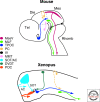
Figure 1.
Figure 1.
Schematic representation of the early axonal scaffold in mouse and Xenopus. Abbreviations: MesV, descending tract of the mesencephalic nucleus of the trigeminal nerve; MLF, medial longitudinal fasciculus; TPOC, tract of the postoptic commissure; PC, posterior commissure; IV, trochlear nerve; MMT, mammilothalamic tract; SOT, supraoptic tract; AC, anterior commissure; DVDT, dorsoventral diencephalic tract; POC, postoptic commissure; mes, mesencephalon; die, diencephalon; tel, telencephalon; rhomb, rhombencephalon.
Figure 2.
Figure 2.
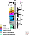
Flat-mount view of the HH21 chick rhombencephalon illustrating its early segmentation into 8 rhombomeres (r1-r8). The hox code specific for each rhombomere or odd- and even-numbered pair of rhombomeres is indicated by a color code. The different cranial motornuclei and nerve root are also represented. Abbreviations: IV, trochlear nucleus; V, trigeminal nucleus; VI, abducens nucleus; VII, facial nucleus; IX, glossopharyngeal nucleus; X, vagus nucleus; XII, hypoglossus nucleus; MHB midbrain hindbrain boundary; Mes, mesencephalon; FP, floorplate. Adapted from Kiecker and Lumsden, 2005.
Figure 3.
Figure 3.
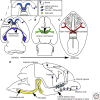
Commissural and longitudinal projections in the forebrain. Both glial and neuronal structures are associated with axonal tracts in the brain. (A–C) depict commissural tracts in schematics of horizontal sections from dorsal to ventral. (A and B) are schematics of the brain, whereas C is a ventral view of the head. Associated with the corpus callosum (blue tract in A) are the glial wedge and indusium griseum glia and the sling cells. Glia are also associated with the hippocampal commissure (purple tract in A) the anterior commissure (green tract in B) and the optic chiasm (red crossing fibers in C). In (D), longitudinal tracts are shown, including the corticothalamic, thalamocortical, cortico-collicular and corticospinal tacts that all pass through the internal capsule. Associated with the internal capsule are the corridor cells. The lateral olfactory tract (LOT) is also shown in D, together with the LOT cells. All schematics are of sections of mouse brain or head at embryonic day 18.
Figure 4.
Figure 4.
Development of dopaminergic projections in the mouse embryo. Dopaminergic axons originate from the substantia nigra (SN) and ventral tegmental area (VTA) in the midbrain and innervate the striatum and cortex. (A) and (B) show the development of this tract at E11 and P0, respectively. They grow rostrally under the repulsive action of Sema3F secreted from the midbrain/hindbrain boundary (MHB, C; C is an enlargement of the boxed area in [A]). The gradient of Sema3F is controlled by Fgf8. Secreted repellents from the mesencephalon and diencephalon/thalamus (B, C) maintain the dopaminergic axons ventrally, whereas factors secreted from the striatum attract them. (D) Dopaminergic axons mostly project ipsilaterally and are maintained away from the midline by Slits and other repellents. Abbreviations: os, optic stalk; tel, telencephalon; die, diencephalon; mes, mesencephalon; rhomb, rhombencephalon; Stri, striatum; Thal, thalamus. Modified from Yamauchi et al., 2009 and Van den Heuvel and Pasterkamp, 2008.
Figure 5.
Figure 5.
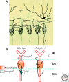
Development of basket cell axons. (A) original drawing by Santiago Ramon y Cajal of a cerebellar basket cell (B) labeled by Golgi staining demonstrating its characteristic axonal arbors (a), the “pinceaux” formations, around the Purkinje cell body and axon. (B) Basket cell axons synapse preferentially on the Purkinje cell axon initial segment (AIS) under the influence of a gradient of Neurofascin 186, stabilized by Ankyrin G. In Ankyrin G knockout mice, the gradient of Neurofascin is abolished and basket cell axons do not synapse preferentially on the AIS. Abbreviations: ML, molecular layer; PCL, Purkinke cell layer; GCL, granule cell layer. A, Cajal drawing. Original conserved at the Instituto Cajal (CSIC), Madrid (Spain). B is adapted from Huang et al., 2007.
Figure 6.
Figure 6.
Serotonin influences axonal arborization during development. In layer IV of the somatosensory cortex, thalamic axons conveying sensory information from the same whisker cluster and arborize in the same domain called a “barrel.” When the level of serotonin is increased during development, such as occurs in MaoA and SERT knockout mice, the barrel field does not form and thalamic axon terminals corresponding to distinct whiskers overlap. Adapted from Gaspar et al., 2003.
Figure 7.
Figure 7.
Advances in techniques for labeling axon tracts and circuits. (A) Carbocyanine dye labeling in the developing brain. DiI-labeled callosal axons are shown in red (arrow in A). Labeling was performed on fixed embryonic day 17 mouse brain. (image courtesy of Dr Celine Plachez, University of Maryland). (B) Labeling of neurons in the brain of a “brainbow” mouse. Different neurons are visualized with different hues of color generated by Cre/loxP recombination in transgenic mice (image courtesy of Dr Jeff Lichtman and Dr Tamily Weissman, Harvard University). (C) Diffusion-weighted (30 directions) magnetic resonance image acquired at 16.4 Tesla—colormap demonstrating commissural tracts in a midsagittal view. Based on their orientation, commissural fibers have been color-coded in red, including the corpus callosum (arrow in C) and anterior commissure (arrowhead in C). (D and E) are tractography images of high angular resolution imaging (HARDI/q-ball). In D, regions of interest (ROI) were selected across the brain, with axon tracts shown that pass through the midline. E demonstrates a more selective placement of ROI’s, one at the midline within the anterior commissure (arrowheads in E depict both the anterior and posterior arms of the anterior commissure that pass through the ROI at the midline), and one in the hindbrain at the midline within the middle cerebellar peduncle and pontine transverse fibers (arrowhead in E). Images in C–E courtesy of Dr Nyoman Kurniawan and Dr Randal Moldrich (The University of Queensland). Scale bar in E = 400 µm in A, 80 µm in B, 2 mm in C and E and 1.35 mm in D.

Similar articles

Cited by

References

    1. Alcamo EA, Chirivella L, Dautzenberg M, Dobreva G, Farinas I, Grosschedl R, McConnell SK 2008. Satb2 regulates callosal projection neuron identity in the developing cerebral cortex. Neuron 57:364–377 - PubMed
    1. Andrews GL, Mastick GS 2003. R-cadherin is a Pax6-regulated, growth-promoting cue for pioneer axons. J Neurosci 23:9873–9880 - PMC - PubMed
    1. Ango F, di Cristo G, Higashiyama H, Bennett V, Wu P, Huang ZJ 2004. Ankyrin-based subcellular gradient of neurofascin, an immunoglobulin family protein, directs GABAergic innervation at purkinje axon initial segment. Cell 119:257–272 - PubMed
    1. Ango F, Wu C, Van der Want JJ, Wu P, Schachner M, Huang ZJ 2008. Bergmann glia and the recognition molecule CHL1 organize GABAergic axons and direct innervation of Purkinje cell dendrites. PLoS Biol 6:e103. - PMC - PubMed
    1. Arlotta P, Molyneaux BJ, Jabaudon D, Yoshida Y, Macklis JD 2008. Ctip2 controls the differentiation of medium spiny neurons and the establishment of the cellular architecture of the striatum. J Neurosci 28:622–632 - PMC - PubMed

Publication types

LinkOut - more resources