

A toxin-based probe reveals cytoplasmic exposure of Golgi sphingomyelin
- PMID: 20463009
- PMCID: PMC2903383
- DOI: 10.1074/jbc.M110.105122
A toxin-based probe reveals cytoplasmic exposure of Golgi sphingomyelin
Abstract
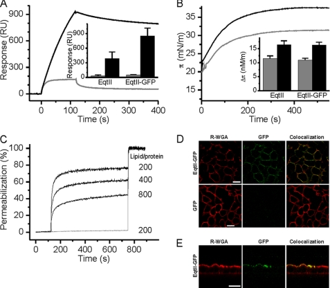
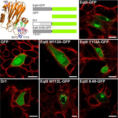
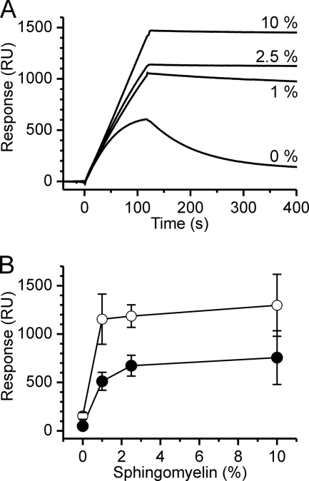
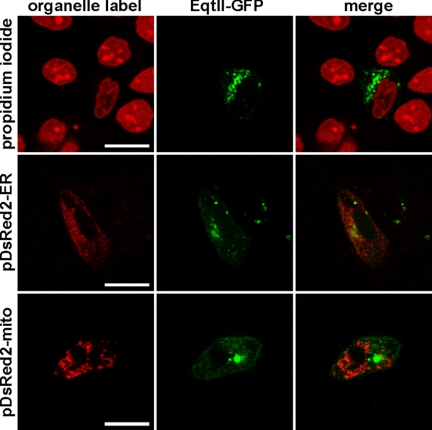
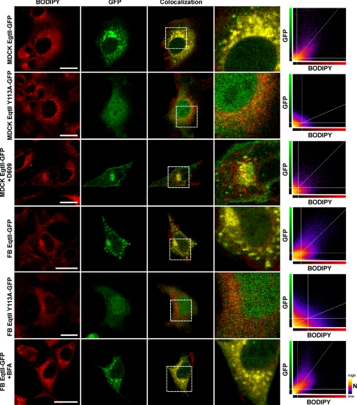
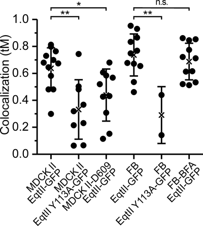
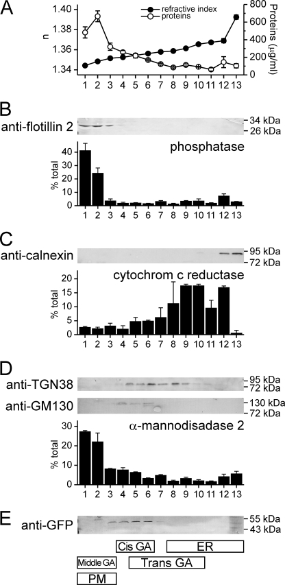
Although sphingomyelin is an important cellular lipid, its subcellular distribution is not precisely known. Here we use a sea anemone cytolysin, equinatoxin II (EqtII), which specifically binds sphingomyelin, as a new marker to detect cellular sphingomyelin. A purified fusion protein composed of EqtII and green fluorescent protein (EqtII-GFP) binds to the SM rich apical membrane of Madin-Darby canine kidney (MDCK) II cells when added exogenously, but not to the SM-free basolateral membrane. When expressed intracellularly within MDCK II cells, EqtII-GFP colocalizes with markers for Golgi apparatus and not with those for nucleus, mitochondria, endoplasmic reticulum or plasma membrane. Colocalization with the Golgi apparatus was confirmed by also using NIH 3T3 fibroblasts. Moreover, EqtII-GFP was enriched in cis-Golgi compartments isolated by gradient ultracentrifugation. The data reveal that EqtII-GFP is a sensitive probe for membrane sphingomyelin, which provides new information on cytosolic exposure, essential to understand its diverse physiological roles.
Figures
Similar articles
-
A non-toxic equinatoxin-II reveals the dynamics and distribution of sphingomyelin in the cytosolic leaflet of the plasma membrane.Sci Rep. 2024 Jul 23;14(1):16872. doi: 10.1038/s41598-024-67803-2. Sci Rep. 2024. PMID: 39043900 Free PMC article.
-
Molecular determinants of sphingomyelin specificity of a eukaryotic pore-forming toxin.J Biol Chem. 2008 Jul 4;283(27):18665-77. doi: 10.1074/jbc.M708747200. Epub 2008 Apr 28. J Biol Chem. 2008. PMID: 18442982
-
Neutron reflection study of the interaction of the eukaryotic pore-forming actinoporin equinatoxin II with lipid membranes reveals intermediate states in pore formation.Biochim Biophys Acta. 2016 Apr;1858(4):640-52. doi: 10.1016/j.bbamem.2015.12.019. Epub 2015 Dec 17. Biochim Biophys Acta. 2016. PMID: 26706098
-
Imaging local sphingomyelin-rich domains in the plasma membrane using specific probes and advanced microscopy.Biochim Biophys Acta. 2014 May;1841(5):720-6. doi: 10.1016/j.bbalip.2013.07.003. Epub 2013 Jul 13. Biochim Biophys Acta. 2014. PMID: 23860017 Review.
-
Molecular mechanism of sphingomyelin-specific membrane binding and pore formation by actinoporins.Adv Exp Med Biol. 2010;677:106-15. Adv Exp Med Biol. 2010. PMID: 20687484 Review.
Cited by
-
Characterization of the Lipid-Binding Site of Equinatoxin II by NMR and Molecular Dynamics Simulation.Biophys J. 2015 Apr 21;108(8):1987-96. doi: 10.1016/j.bpj.2015.03.024. Biophys J. 2015. PMID: 25902438 Free PMC article.
-
Ceramide-induced apoptosis in renal tubular cells: a role of mitochondria and sphingosine-1-phoshate.Int J Mol Sci. 2015 Mar 5;16(3):5076-124. doi: 10.3390/ijms16035076. Int J Mol Sci. 2015. PMID: 25751724 Free PMC article. Review.
-
Imaging the lipid-phase-dependent pore formation of equinatoxin II in droplet interface bilayers.Biophys J. 2014 Apr 15;106(8):1630-7. doi: 10.1016/j.bpj.2013.11.4507. Biophys J. 2014. PMID: 24739162 Free PMC article.
-
Dynamic transbilayer lipid asymmetry.Cold Spring Harb Perspect Biol. 2011 May 1;3(5):a004671. doi: 10.1101/cshperspect.a004671. Cold Spring Harb Perspect Biol. 2011. PMID: 21436058 Free PMC article. Review.
-
Perfringolysin O Theta Toxin as a Tool to Monitor the Distribution and Inhomogeneity of Cholesterol in Cellular Membranes.Toxins (Basel). 2016 Mar 8;8(3):67. doi: 10.3390/toxins8030067. Toxins (Basel). 2016. PMID: 27005662 Free PMC article. Review.
References
Publication types
MeSH terms
Substances
LinkOut - more resources
Full Text Sources
Other Literature Sources
