. 2010 Jul 16;285(29):22542-53.
doi: 10.1074/jbc.M109.094557. Epub 2010 May 12.

Molecular consequences of the ACVR1(R206H) mutation of fibrodysplasia ossificans progressiva

Affiliations

Molecular consequences of the ACVR1(R206H) mutation of fibrodysplasia ossificans progressiva

Gin-Ah Song et al. J Biol Chem. .

Abstract

Fibrodysplasia ossificans progressiva (FOP), a rare genetic and catastrophic disorder characterized by progressive heterotopic ossification, is caused by a point mutation, c.617G>A; p.R206H, in the activin A receptor type 1 (ACVR1) gene, one of the bone morphogenetic protein type I receptors (BMPR-Is). Although altered BMP signaling has been suggested to explain the pathogenesis, the molecular consequences of this mutation are still elusive. Here we studied the impact of ACVR1 R206H mutation on BMP signaling and its downstream signaling cascades in murine myogenic C2C12 cells and HEK 293 cells. We found that ACVR1 was the most abundant of the BMPR-Is expressed in mesenchymal cells but its contribution to osteogenic BMP signal transduction was minor. The R206H mutant caused weak activation of the BMP signaling pathway, unlike the Q207D mutant, a strong and constitutively active form. The R206H mutant showed a decreased binding affinity for FKBP1A/FKBP12, a known safeguard molecule against the leakage of transforming growth factor (TGF)-beta or BMP signaling. The decreased binding affinity of FKBP1A to the mutant R206H ACVR1 resulted in leaky activation of the BMP signal, and moreover, it decreased steady-state R206H ACVR1 protein levels. Interestingly, while WT ACVR1 and FKBP1A were broadly distributed in plasma membrane and cytoplasm without BMP-2 stimulation and then localized in plasma membrane on BMP-2 stimulation, R206H ACVR1 and FKBP1A were mainly distributed in plasma membrane regardless of BMP-2 stimulation. The impaired binding to FKBP1A and an altered subcellular distribution by R206H ACVR1 mutation may result in mild activation of osteogenic BMP-signaling in extraskeletal sites such as muscle, which eventually lead to delayed and progressive ectopic bone formation in FOP patients.


Figures
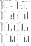
FIGURE 1.
FIGURE 1.
ACVR1 is a strongly expressed BMP type I receptor but plays a minor role in BMP signal transduction in myogenic C2C12 cells. A, after visual confluence of seeded C2C12 cells, the cells were cultured for an additional 24 h with, or without, rhBMP-2 (30 ng/ml). Total RNA was harvested, and mRNA levels of BMP receptors were analyzed by RT-qPCR. (**, p < 0.001 versus Bmpr1a expression in the untreated condition.) B, C2C12 cells were transiently transfected with ACVR1 or empty vector, and the transfected cells were cultured for an additional 24 h with, or without, rhBMP-2 (30 ng/ml). The mRNA levels of the BMP downstream genes, Dlx5 and Alp, were determined by RT-qPCR. (**, p < 0.001, *p < 0.01 versus untreated vehicle.) C, C2C12 cells were transfected with a control siRNA (si-control), Bmpr1a (si-Bmpr1a), and ACVR1 (si-Acvr1). Cells were cultured for 24–36 h and treated with, or without, rhBMP-2 (30 ng/ml) for an additional 24 h. (**, p < 0.001; *, p < 0.01 versus untreated Si-control.) D, C2C12 cells were transfected with the siRNAs with, or without, ACVR1 overexpression. After reaching visual confluence, cells were cultured for an additional 24 h with, or without, rhBMP-2 (30 ng/ml). (Data represent mean ± S.D. Statistical differences compared with the respective controls are depicted by **, p < 0.001; *, p < 0.01 by the two-tailed Student's t test. ns, not significant.)
FIGURE 2.
FIGURE 2.
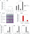
ACVR1R206H is a weak activating mutant unlike ACVR1Q207D, a constitutively active mutant. A, C2C12 cells were transiently transfected with ACVR1 WT, R206H mutant, dominant negative mutant (K235R), or constitutively active mutant (Q207D). After 24 h of transfection, cells were treated with, or without, rhBMP-2 (30 ng/ml) for an additional 24 h. The Alp mRNA level was determined by RT-qPCR. (**, p < 0.001; *, p < 0.01 versus untreated WT.) B, C2C12 cells were transiently transfected with four different expression constructs of ACVR1 or Smad5. After 24 h of transfection, cells were treated with 50 ng/ml rhBMP-2, 500 nm FK506, or a combination of both agents for additional 24 h. Then after a further 3 days of culture, cells were fixed, and ALP activity was cytochemically determined. C, C2C12 cells were co-transfected with a control siRNA (si-control) or si-Smad1 plus si-Smad5 with ACVR1 WT or R206H overexpression. Cells were cultured for 24–36 h, and the mRNA levels of the BMP downstream genes, Dlx5, were determined by RT-qPCR (*, p < 0.01 versus WT with si-control.) D, C2C12 cells, stably transfected with ACVR1 WT or R206H, were cultured. After reaching visual confluence, cells were cultured for an additional 24 h with, or without, rhBMP-2 (30 ng/ml). The mRNA levels of BMP downstream genes (Dlx5, Alp, and Msx2) were determined by RT-qPCR. (**, p < 0.001; *, p < 0.01 versus untreated WT by the two-tailed Student's t test. ns, not significant.)
FIGURE 3.
FIGURE 3.
Reduced and impaired FKBP1A binding for ACVR1R206H results in leakage of BMP signaling. A, C2C12 cells, stably transfected with ACVR1 WT or R206H, were then transfected with FKBP1A. After 24 h of transfection, the cells were treated with 50 ng/ml rhBMP-2 or 1 μm FK506 for an additional 24 h. ALP activity was cytochemically determined as for Fig. 2B. B, cells described in A were harvested after treatment with rhBMP-2 or FK506 for 24 h. The Alp mRNA level was analyzed by RT-qPCR. (**, p < 0.001; *, p < 0.01 versus untreated WT by the two-tailed Student's t test.) C, C2C12 cells, stably transfected with ACVR1 WT or R206H, were treated with 50 ng/ml of rhBMP-2 or 500 nm FK506 for 24 h and Alp activity was determined cytochemically. D, whole cell lysates of HEK293 cells transfected with the four ACVR1 expression constructs (WT, R206H, K235R, Q207D) were immunoprecipitated by α-Myc (pcDNA4-FKBP1A-myc) and then immunoblotted with α-V5 (pcDNA6-ACVR1-V5) or α-Myc antibody. The intensities of the immunoprecipitated ACVR1 bands were normalized to respective FKBP1A band using the average intensity of the WT as 100% from two independent experiments. Data represent the means ± S.D.
FIGURE 4.
FIGURE 4.
ACVR1-FKBP1A interaction stabilizes ACVR1 protein, ACVR1R206H mutation causes reduced amount of protein because of reduced affinity for FKBP1A. A, lysates of HEK293 cells, transfected with ACVR1 WT, or R206H, were analyzed by SDS/PAGE and Western blotting with the α-V5 (ACVR1) antibody. The intensities of the ACVR1 bands were normalized to the actin bands using the average intensity of the WT as 100% from three independent experiments. B, HEK293 cells were transfected with the four ACVR1 constructs with, or without FKBP1A, and then analyzed by Western blotting with the α-V5 or α-Myc antibodies. C, stable C2C12 cells carrying WT or R206H were analyzed by Western blotting. D, RT-PCR analysis of the mRNA levels of overexpressed Acvr1 WT and R206H. Forward and reverse primers were designed to include the V5 and BGH reverse region, which are distal sequences from ACVR1 ORF insert in pcDNA6-ACVR1-v5 plasmids. Plasmid and RNA without reverse transcription, as a template, was included for a positive and negative control reaction, respectively.
FIGURE 5.
FIGURE 5.
The ACVR1R206H mutation shows a different subcellular distribution to WT by quantitative analysis. A and B, FKBP1A with WT or R206H were transiently transfected in HEK293 cells with an increasing ratio of FKBP1A plasmid to ACVR1 plasmid as indicated (FKBP1A relative ratio). After 24 h, whole cell lysates were harvested using TNE-CHAPS lysis buffer to solubilize transmembrane receptors or TNE lysis buffer to collect total soluble cytosolic proteins. The graphs in the lower panel represent the densitometric analysis of the Western blots of the upper panel. Each experiment was repeated at least twice.
FIGURE 6.
FIGURE 6.
Different subcellular distribution between WT and ACVR1R206H. A, C2C12 stable cells carrying WT ACVR1 or R206H were transiently transfected with FKBP1A and then immunostained with α-V5 (green: ACVR1) and α-Myc (RED: FKBP1A) antibody followed by Alexa F488-conjugated anti-mouse and Qdot 655-conjugated anti-rabbit secondary antibodies. The view of a confocal slice midway through the thickness of the cells was taken. WT shows a broadly dispersed cytosolic pattern with FKBP1A. (Long thin white arrow, ER; short thick white arrow, Golgi apparatus.) B, after 30 min of treatment with 50 ng/ml rhBMP-2, the cells were fixed and immunostained. The view of a confocal slice was taken midway through the height of the cells. The subcellular distribution of WT with rhBMP-2 treatment, and the R206H mutant without rhBMP-2 treatment was compared. C, serial confocal slices were taken in the upper and lower 1 to 3 μm from the midway through the height of the cells. (Blue, DAPI; yellow, merged area; scale bars: 10 μm or 20 μm.)

References

    1. Connor J. M., Evans D. A. (1982) J. Bone Joint. Surg. Br. 64, 76–83 - PubMed
    1. Connor J. M., Skirton H., Lunt P. W. (1993) J. Med. Genet. 30, 687–689 - PMC - PubMed
    1. Shafritz A. B., Shore E. M., Gannon F. H., Zasloff M. A., Taub R., Muenke M., Kaplan F. S. (1996) N. Engl. J. Med. 335, 555–561 - PubMed
    1. Cohen R. B., Hahn G. V., Tabas J. A., Peeper J., Levitz C. L., Sando A., Sando N., Zasloff M., Kaplan F. S. (1993) J. Bone Joint. Surg. Am. 75, 215–219 - PubMed
    1. Kitterman J. A., Kantanie S., Rocke D. M., Kaplan F. S. (2005) Pediatrics 116, e654–661 - PubMed

Publication types

Substances